

Review
doi: 10.1002/iub.53.
Electron crystallography of aquaporins
Affiliations
- PMID: 18465794
- PMCID: PMC2690706
- DOI: 10.1002/iub.53
Item in Clipboard
Review
Electron crystallography of aquaporins
IUBMB Life.
2008 Jul.
Abstract
Aquaporins are a family of ubiquitous membrane proteins that form a pore for the permeation of water. Both electron and X-ray crystallography played major roles in determining the atomic structures of a number of aquaporins. This review focuses on electron crystallography, and its contribution to the field of aquaporin biology. We briefly discuss electron crystallography and the two-dimensional crystallization process. We describe features of aquaporins common to both electron and X-ray crystallographic structures; as well as some structural insights unique to electron crystallography, including aquaporin junction formation and lipid-protein interactions.
Figures

Structure of aquaporin-1. A: Aquaporins are tetramers but each monomer forms a functional water pore (asterisk). B: Side view of an aquaporin-1 monomer. Six transmembrane helices pack against one another forming a barrel-like structure with the hydrophilic water pore at the center (green). The ar/R constriction site, or selectivity filter, is shown in red (Protein Data Bank accession No. 1FQY).
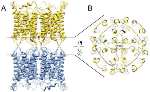
Structure of the aquaporin-0 mediated membrane junction. A: Side view of the aquaporin-0 junction. Two opposing tetramers interact in a head-to-head fashion. B: A major adhesive contact is mediated by proline 38, forming a rosette-like structure at the very center of the stacked tetramers (Protein Data Bank accession No. 2B6O).
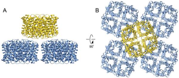
Double layered aquaporin-4 2D crystals. A and B: Side and top views of the double-layered 2D crystals of aquaporin-4, respectively. Each aquaporin-4 tetramer contacts four tetramers from the opposing layer (Protein Data Bank accession No. 2D57).
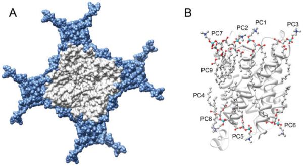
Lipid-protein interactions in aquaporin-0 double-layered 2D crystals. A: Lipids (blue) pack tightly around the aquaporin-0 tetramer (grey). Lipid acyl chains fit snuggly into irregularities on the protein surface. B: The nine unique DMPC lipid molecules (PC1-9) identified per aquaporin-0 monomer. PC1-7 are annular lipids while PC8 and 9 represent bulk lipids (Protein Data Bank accession No. 2B6O).
References
-
- Agre P, Sasaki S, Chrispeels MJ. Aquaporins: a family of water channel proteins. Am. J. Physiol. 1993;265:F461. - PubMed
-
- Agre P, Kozono D. Aquaporin water channels: molecular mechanisms for human diseases. FEBS Lett. 2003;555:72–78. - PubMed
-
- Yang B, Verkman AS. Water and glycerol permeabilities of aquaporins 1-5 and MIP determined quantitatively by expression of epitope-tagged constructs in Xenopus oocytes. J. Biol. Chem. 1997;272:16140–16146. - PubMed
-
- Gonen T, Walz T. The structure of aquaporins. Q. Rev. Biophys. 2006;39:361–396. - PubMed
-
- Gyobu N, Tani K, Hiroaki Y, Kamegawa A, Mitsuoka K, Fujiyoshi Y. Improved specimen preparation for cryo-electron microscopy using a symmetric carbon sandwich technique. J. Struct. Biol. 2004;146:325–333. - PubMed
Publication types
MeSH terms
Substances
Grants and funding
LinkOut - more resources
Full Text Sources
