. 2008 Oct 1;101(2):345-56.
doi: 10.1002/bit.21911.

A new technique for primary hepatocyte expansion in vitro

Affiliations

A new technique for primary hepatocyte expansion in vitro

Cheul H Cho et al. Biotechnol Bioeng. .

Abstract

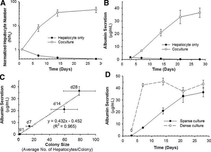
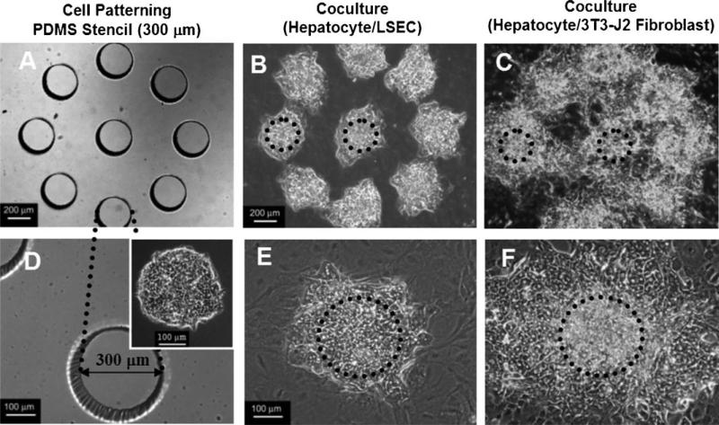
The current application for many potential cell-based treatments for liver failure is limited by the low availability of mature functional hepatocytes. Although adult hepatocytes have a remarkable ability to proliferate in vivo, attempts to proliferate adult hepatocytes in vitro have been less successful. In this study, we investigated the effect of coculture cell type on the proliferative response and the functional activities of hepatocytes. We show, for the first time, a robust proliferative response of primary adult rat hepatocytes when cocultured with mouse 3T3-J2 fibroblasts. Hepatocytes cultured at low density on growth-arrested 3T3-J2 fibroblast feeder layers underwent significantly higher proliferation rates than when cultured on feeder layers made of four other cell types. Increasing colony size correlated with an increase in hepatocellular functions. The proliferating hepatocytes retained their morphologic, phenotypic, and functional characteristics. Using a cell patterning technique, we found that 3T3-J2 fibroblasts stimulate DNA synthesis in hepatocytes by short-range heterotypic cell-cell interactions. When hepatocytes that proliferated in cocultures were harvested and further subcultured either on 3T3-J2 fibroblast feeders or in the collagen sandwich configuration, their behavior was similar to that of freshly isolated hepatocytes. We conclude that adult rat hepatocytes can proliferate in vitro in a coculture cell type-dependent manner, and can be serially propagated by coculturing with 3T3-J2 fibroblasts while maintaining their differentiated characteristics. Our results also suggest that one of the major reasons for the functional differences in hepatocyte cocultures may be due to the different proliferative responses of hepatocytes as a function of coculture cell type. This study provides new insights in the roles of coculture cell types and cell-cell interactions in the modulation of hepatic proliferation and function.


Figures

Figure 1
Figure 1
Morphologic and phenotypic characteristics of hepatocytes cultured alone (A) and cocultured on growth arrested 3T3-J2 fibroblast feeder layers (B–D). Hepatocytes were sparsely seeded (1.25 × 10³ cells/cm2) to better visualize individual hepatocyte colonies. A: Morphology, F-actin distribution, and albumin staining in hepatocytes cultured alone after 6 days of culture. B: Phase contrast images of the same single hepatocyte colony observed after 1, 6, and 14 days of culture in hepatocyte/3T3-J2 fibroblast coculture. Arrow points to hepatocyte colony 1 day after seeding. The dashed circles indicate the morphology of proliferating hepatocytes captured at the same location during culture periods. Scale bar, 100 μm. C: Albumin (mature hepatocyte marker) staining and BrdU (DNA synthesis marker) uptake after 8, 14, and 25 days of culture. Scale bar, 100 μm. D: Immunofluorescence and immunohistochemical analysis for actin filament distribution, albumin synthesis, and glycogen storage on day 27.
Figure 2
Figure 2
The proliferation rate and function of hepatocytes cultured on growth arrested 3T3-J2 fibroblast feeder layers. Hepatocytes cultured alone on collagen coated dishes served as controls. A: The normalized hepatocyte number (N/No) over time in sparse culture (1.25 × 10³ cells/cm2). The hepatocyte numbers (N) were normalized to the initial number of hepatocytes (No). B: Albumin secretion over time in sparse culture. C: Correlation between hepatocyte colony sizes and albumin secretion. D: Albumin secretion as a function of time for sparse (1.25 × 10³ cells/cm2) and dense (15.63 × 10³ cells/cm2) culture. Data shown are means ± SD of three independent experiments in duplicate.
Figure 3
Figure 3
Role of heterotypic interface on hepatocyte DNA synthesis and albumin expression. Distribution of DNA synthetic activity, as reported by BrdU uptake, and albumin staining in a 1,000 μm diameter hepatocyte island (A–F) and in a doughnut-shape hepatocyte island (G–L). Scale bar, 500 μm (B,C and H,I) and 100 μm (D–F and J–L).
Figure 4
Figure 4
Effect of feeder layer cell types on hepatocyte proliferation and function. 3T3-J2, NIH-3T3, MEF, LSEC, and MVEC were used as feeder layers. A: Typical hepatocyte colony size and albumin staining in cocultures with the various feeder cell types on day 14. B: Average number of hepatocytes per colony in cocultures on day 14. C: Normalized hepatocyte number (N/No) in cocultures on day 14. D,E: Albumin secretion (D) and urea synthesis (E) by the hepatocytes cocultured with the various cell types on day 14. F,G: Correlation between hepatocyte colony sizes and liver-specific functions (albumin secretion and urea synthesis) in cocultures with the various feeder cell types on day 14. Data shown are means ± SD of two independent experiments in duplicate. *P < 0.05; **P < 0.005. Hepatocytes were seeded in sparse culture (1.25 × 10³ cells/cm2) on all feeder layers. [Color figure can be seen in the online version of this article, available at www.interscience.wiley.com.]
Figure 5
Figure 5
Comparison of hepatocyte proliferation between micropatterned cocultures on 3T3-J2 fibroblasts versus LSEC feeder layers. Hepatocytes were seeded as 300 μm diameter islands using PDMS stencils, and observed after 13 days of culture. A,D: Microfabricated PDMS stencil with 300 μm diameter holes. Inset in panel D shows the micropatterned hepatocytes on day 1. B,E: Hepatocyte and LSEC coculture on day 13. C,F: Hepatocyte and 3T3-J2 fibroblast coculture on day 13. Dashed circle lines indicate the original island size. Scale bar, 200 μm (upper panels) and 100 μm (lower panels).
Figure 6
Figure 6
Morphology and function of hepatocytes in subcultures. Hepatocytes were cocultured with 3T3-J2 fibroblasts for 35 days, detached from the substrate by dispase/collagenase treatment, dispersed in a single cell suspension by trypsin digestion, replated, and subcultured for another 30 days. Replating was either at high density (~125 × 103 cells/cm2) in a collagen sandwich configuration, or at low density (~1.25 × 10³ cells/cm2) on growth arrested 3T3-J2 fibroblast feeder layers. A: Albumin secretion in the subcultures. B,C: Morphology of hepatocytes in the subcultures. Scale bar, 100 μm. D: Morphology of hepatocytes cocultured on 3T3-J2 fibroblast feeder layer in the subculture on day 39 (4 days after replating), day 47 (12 days after replating), and day 63 (28 days after replating), and double staining for albumin expression and BrdU incorporation on day 63 (28 days after replating). Scale bar, 100 μm.

References

    1. Asahina K, Fujimori H, Shimizu-Saito K, Kumashiro Y, Okamura K, Tanaka Y, Teramoto K, Arii S, Teraoka H. Expression of the liver-specific gene Cyp7a1 reveals hepatic differentiation in embryoid bodies derived from mouse embryonic stem cells. Genes Cells. 2004;9(12):1297–1308. - PubMed
    1. Bhatia SN, Balis UJ, Yarmush ML, Toner M. Probing heterotypic cell interactions: Hepatocyte function in microfabricated co-cultures. J Biomater Sci Polym Ed. 1998;9(11):1137–1160. - PubMed
    1. Bhatia SN, Balis UJ, Yarmush ML, Toner M. Effect of cell-cell interactions in preservation of cellular phenotype: Cocultivation of hepatocytes and nonparenchymal cells. FASEB J. 1999;13(14):1883–1900. - PubMed
    1. Bissell DM, Arenson DM, Maher JJ, Roll FJ. Support of cultured hepatocytes by a laminin-rich gel. Evidence for a functionally significant subendothelial matrix in normal rat liver. J Clin Invest. 1987;79(3):801–812. - PMC - PubMed
    1. Block GD, Locker J, Bowen WC, Petersen BE, Katyal S, Strom SC, Riley T, Howard TA, Michalopoulos GK. Population expansion, clonal growth, and specific differentiation patterns in primary cultures of hepatocytes induced by HGF/SF, EGF and TGF alpha in a chemically defined (HGM) medium. J Cell Biol. 1996;132(6):1133–1149. - PMC - PubMed

Publication types

LinkOut - more resources