. 2008 Oct;23(10):1619-28.
doi: 10.1359/jbmr.080502.

SMAD3 functions as a transcriptional repressor of acid-sensing ion channel 3 (ASIC3) in nucleus pulposus cells of the intervertebral disc

Affiliations

SMAD3 functions as a transcriptional repressor of acid-sensing ion channel 3 (ASIC3) in nucleus pulposus cells of the intervertebral disc

Yoshiyasu Uchiyama et al. J Bone Miner Res. 2008 Oct.

Abstract

The goal of this investigation was to study the regulation of acid-sensing ion channel (ASIC)3 expression by TGFbeta in the nucleus pulposus cells of the intervertebral disc. Analysis of human nucleus pulposus tissue indicated decreased ASIC3 and elevated TGFbeta expression in the degenerate state. In a parallel study, treatment of nucleus pulposus cells with TGFbeta resulted in decreased expression of ASIC3 mRNA and protein. Suppression of ASIC3 promoter activity was evident when the nucleus pulposus cells were treated with TGFbeta or co-transfected with the constitutively active ALK5 or a smad3 construct. On the other hand, co-transfection of dominant negative smad3 or smad7 restored ASIC3 promoter activity. We validated the role of smad3 in controlling ASIC3 expression using cells derived from smad3-null mice. ASIC3 promoter activity in the null cells was 2- to 3-fold higher than the wildtype cells. Moreover, expression of smad3 in null cells decreased ASIC3 promoter activity by almost 50%. Further studies using deletion constructs and trichostatin A treatment showed that the full-length smad3 was necessary, and the suppression involved recruitment of histone deacetylase to the promoter. To determine the mechanism, we evaluated the rat ASIC3 promoter sequence and noted the presence of two smad interacting CAGA box motifs. Gel-shift and supershift analysis indicated that smad3 protein was bound to this motif. Chromatin immunoprecipitation analysis confirmed that smad3 bound both the CAGA elements. Results of these studies clearly show that TGFbeta is highly expressed in the degenerate disc and through smad3 serves as a negative regulator of ASIC3 expression.


Figures

FIG. 1
FIG. 1
Effect of TGFβ on ASIC-3 expression in nucleus pulposus cells. (A) Immunofluorescent detection of ASIC3 in nucleus pulposus cells. Cells were treated with an antibody to ASIC3, and cell nuclei were stained with propidium iodide (PI). NP cells showed strong expression of ASIC3. Magnification, ×20. (B and C) Cells were treated with TGFβ (1–10 ng/ml) for 24–72 h, and ASIC3 expression was analyzed. (B) Real-time RT-PCR analysis of ASIC3 indicates decreased ASIC3 mRNA expression after treatment with TGFβ. (C) Western blots analysis showed a dose-dependent reduction in ASIC3 protein expression in TGFβ-treated NP cells. (D) Multiple Western blots of ASIC3 quantified by densitometric analysis. Note, NP cells exhibited a suppression of ASIC3 levels when TGFβ was used at a concentration of 10 ng/ml. (E) RT-PCR analysis of normal (N) and Thompson graded (grade 3, G3; and grade 4, G4) degenerate human nucleus pulposus tissue from lumbar discs. Note the high level of expression of ASIC3 in normal tissue that is significantly decreased in degenerate tissue. Degenerate tissue evidenced a higher expression of TGFβ mRNA than normal control tissue. Quantitative data represents mean ± SD of three independent experiments (n = 3); *p < 0.05; ns, nonsignificant.
FIG. 2
FIG. 2
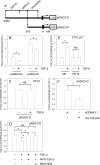
Effect of TGFβ in ASIC3 promoter activity. (A) Cartoon showing map of successive PCR generated 5` deletion constructs of the rat ASIC3 promoter. The ASIC3 promoter contains three distinct domains: proximal, middle, and a distal activating domain. The ASIC3-D construct is a 2925-bp fragment containing 2831 bp of the upstream ASIC3 promoter sequence linked to 94 bp of exon 1 (i.e., −2831 to +94), whereas the ASIC3-P constructs contains a 1065-bp (−971 to + 94) fragment. (B and C) NP or PC12 cells were transfected with ASIC-3 reporter constructs along with pRL-TK vector. Cells were treated with TGFβ (10 ng/ml) for 24 h, and luciferase reporter activity was measured. NP cells showed significant suppression in activities of both ASIC-3 constructs (B). PC12 cells did not show a TGFβ-mediated decrease in pASIC-3D activity (D) NP cells transfected with ASIC3 reporter were treated with anti-TGFβ antibody (10 μg/ml) or SB431542 (10 μM) along with TGFβ3. Both anti-TGFβ antibody and SB431542 reversed the suppressive effects of TGFβ on ASIC3 promoter activity. (E) TGFβ reactivity of NP and PC12 cells assessed by measuring activation of the 3TP-Lux reporter. Note, only NP cells showed induction in 3TP-Lux activity after TGFβ treatment. (F) NP cells were transfected with an ASIC3 reporter along with constitutively active type I TGFßR (ALK5) construct or empty vector pcDNA3. Constitutive ALK5 signaling suppressed ASIC3 promoter activity in NP cells. Values shown are mean ± SD of three independent experiments performed in triplicate. *p < 0.05.
FIG. 3
FIG. 3
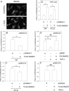
Smad3 regulation of ASIC3 promoter activity. (A) Nucleus pulposus cells were treated with TGFβ for 6 h and probed with anti-Smad3 antibody. Note nuclear accumulation of smad3 protein in treated cells (arrows). (B and C) ASIC3 reporter construct along with full-length Smad3 construct, or empty backbone pcDNA3.1, was transfected into (B) NP cells and (C) PC12 cells, and luciferase activity was measured. Expression of smad3 resulted in significant suppression of ASIC3 promoter activity in both cell types. (D) Transcriptional activity of transfected Smad3 was measured by determining 3TP-Lux reporter activity. Note, smad3 induced 3TP-Lux activation in both NP and PC12 cells. (E) NP cells were transfected with ASIC3 reporter along with DN-Smad3 construct or empty backbone vector pRK5F and treated with TGFβ. Note, expression of DN-Smad3 rescued cells from the suppressive effect of TGFβ on ASIC3 promoter activity. (F) NP cells were transfected with full-length Smad7 construct or empty backbone vector pcDNA3 along with ASIC3 reporter, and luciferase activity was measured after TGFβ treatment. Smad7 expression preserved ASIC3 promoter activity in the presence of TGFβ. Values shown are mean ± SD of three independent experiments performed in triplicate. *p < 0.05.
FIG. 4
FIG. 4
Effect of Smad3 deletion on ASIC-3 promoter activity. (A) Smad3 wildtype (WT: +/+) and null (−/−) MEF were transfected with ASIC-3 reporter constructs along with pRL-TK control vector, and luciferase reporter activity was measured. Null cells showed a significant increase in basal ASIC-3 reporter activity compared with WT cells. (B) Smad3-null cells were transfected with ASIC-3 reporter construct along with full-length Smad3 construct or empty backbone vector pCDNA3.1, and luciferase activity was measured. Forced expression of Smad3 in null cells resulted in a significant suppression in ASIC-3 promoter activity. Values shown are mean ± SD of three independent experiments. *p < 0.05.
FIG. 5
FIG. 5
Wildtype Smad3 is required for regulation of ASIC3 promoter activity. ASIC3 reporter construct along with wildtype (WT)-Smad3 construct or truncated Smad3-encoding plasmids; Smad3NL (MH1 domain and linker) or Smad3C (MH2 domain); or empty vector was transfected into NP cells, and luciferase activity was measured. Expression of WT-Smad3 resulted in suppression, whereas plasmids encoding truncated Smad3 protein did not suppress ASIC3 promoter activity. (B) NP cells were transfected with ASIC3 reporter along with smad3 construct, or empty vector, and treated with trichostatin A (TSA; 100 nM) or vehicle (DMSO) for 24 h. Treatment of TSA restored expression of ASIC3 in the presence of smad3. Values shown are mean ± SD of three independent experiments performed in triplicate. *p < 0.05.
FIG. 6
FIG. 6
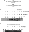
Interaction of Smad3 with ASIC-3 promoter. (A) DNA sequence of the promoter region of the rat ASIC3 gene. The CAGA consensus sequence is marked in bold and underlined. The ASIC3 promoter contains two conserved CAGA motifs at −30 and −2429 bp from the transcription start site. (B) Electromobility shift assay to examine functional binding of smad3 to CAGA motif in the rat ASIC3 gene promoter. An oligonucleotide probe containing the CAGA motif (−30 bp) in the rat ASIC3 promoter was incubated with nuclear extracts from nucleus pulposus cells treated with TGFβ and binding was detected using chemiluminescence. Note, there was induction of binding 6 h after TGFβ treatment. Specificity of binding was confirmed by inclusion of a probe containing mutation in the CAGA site (MT probe) or anti-smad3 antibody in the binding reaction. The binding signal is significantly diminished when a mutant probe was used, whereas a supershift of the band was seen in presence of anti-smad3 antibody. (C) ChIP assay was used to examine the interaction of Smad3 with the ASIC3 promoter. PCR amplification was performed using primers pairs that encompass both distal (Dist)-CAGA and proximal (Prox)-CAGA sequences of the ASIC3 promoter. Note, use of smad3 antibody resulted in generation of PCR amplicons containing both distal HRE and proximal HRE. Detectable binding was observed under basal conditions, which was significantly increased after TGFβ treatment. Use of isotype antibody control did not result in the formation of a PCR product.

References

    1. Hassler O. The human intervertebral disc. A micro-angiographical study on its vascular supply at various ages. Acta Orthop Scand. 1969;40:765–772. - PubMed
    1. Risbud MV, Guttapalli A, Stokes DG, Hawkins D, Danielson KG, Schaer TP, Albert TJ, Shapiro IM. Nucleus pulposus cells express HIF-1alpha under normoxic culture conditions: A metabolic adaptation to the intervertebral disc microenvironment. J Cell Biochem. 2006;98:152–159. - PubMed
    1. Agrawal A, Guttapalli A, Narayan S, Albert TJ, Shapiro IM, Risbud M. Normoxic stabilization of HIF-1alpha drives glycolytic metabolism and regulates aggrecan gene expression in nucleus pulposus cells of the rat intervertebral disk. Am J Physiol Cell Physiol. 2007;293:C621–C631. - PubMed
    1. Grunhagen T, Wilde G, Soukane DM, Shirazi-Adl SA, Urban JP. Nutrient supply and intervertebral disc metabolism. J Bone Joint Surg Am. 2006;88:30–35. - PubMed
    1. Grunder S, Geissler HS, Bassler EL, Ruppersberg JP. A new member of acid-sensing ion channels from pituitary gland. Neuroreport. 2000;11:1607–1611. - PubMed

Publication types