. 2008 Jul;69(1):152-63.
doi: 10.1111/j.1365-2958.2008.06271.x. Epub 2008 May 5.

Targeted deletion of SAP1 abolishes the expression of infectivity factors necessary for successful malaria parasite liver infection

Affiliations
Free PMC article

Targeted deletion of SAP1 abolishes the expression of infectivity factors necessary for successful malaria parasite liver infection

Ahmed S I Aly et al. Mol Microbiol. 2008 Jul.
Free PMC article

Abstract

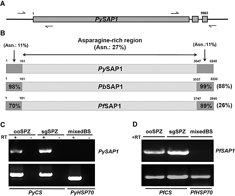
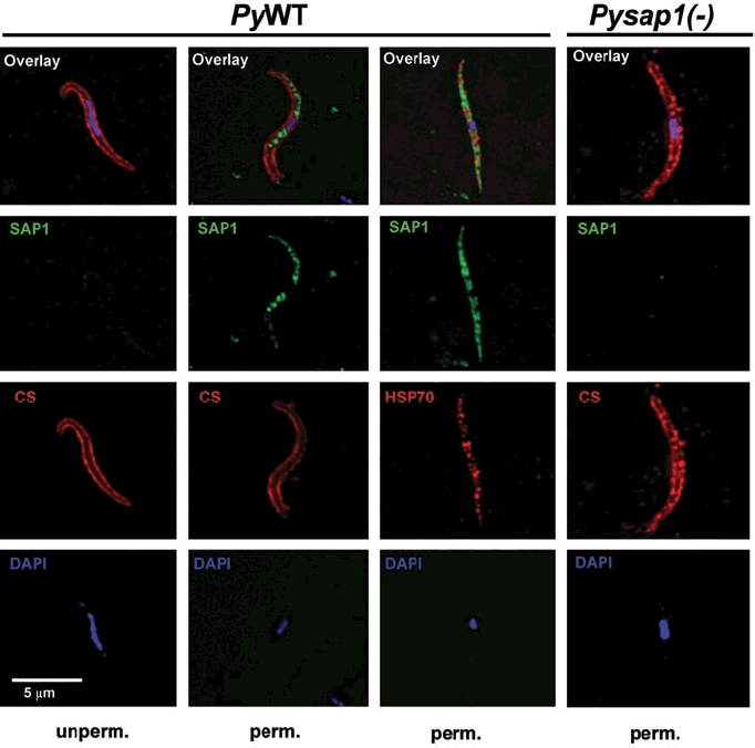
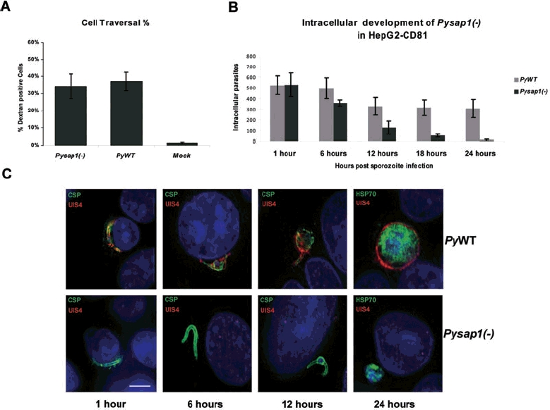
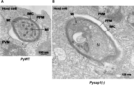
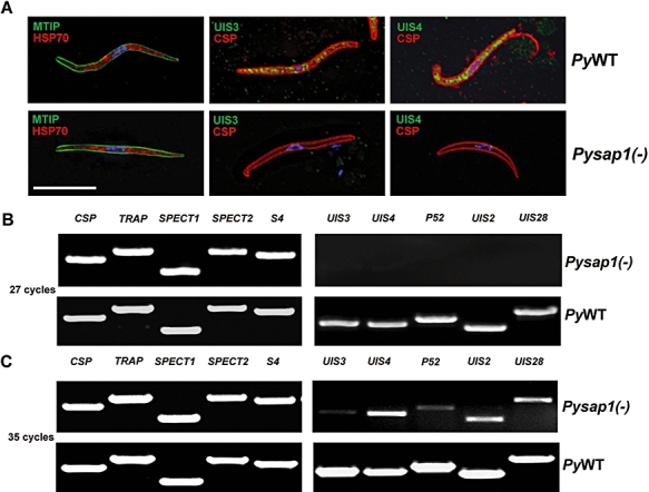
Malaria parasite sporozoites prepare for transmission to a mammalian host by upregulation of UIS (Upregulated in Infectious Sporozoites) genes. A number of UIS gene products are essential for the establishment of the intrahepatocytic niche. However, the factors that regulate the expression of genes involved in gain of infectivity for the liver are unknown. Herein, we show that a conserved Plasmodium sporozoite low-complexity asparagine-rich protein, SAP1 (Sporozoite Asparagine-rich Protein 1), has an essential role in malaria parasite liver infection. Targeted deletion of SAP1 in the rodent malaria parasite Plasmodium yoelii generated mutant parasites that traverse and invade hepatocytes normally but cannot initiate liver-stage development in vitro and in vivo. Moreover, immunizations with Pysap1(-) sporozoites confer long-lasting sterile protection against wild-type sporozoite infection. Strikingly, lack of SAP1 abolished expression of essential UIS genes including UIS3, UIS4 and P52 but not the constitutively expressed genes encoding, among others, sporozoite proteins CSP and TRAP. SAP1 localization to the cell interior but not the nucleus of sporozoites suggests its involvement in a post-transcriptional mechanism of gene expression control. These findings demonstrate that SAP1 is essential for liver infection possibly by functioning as a selective regulator controlling the expression of infectivity-associated parasite effector genes.


Figures

Fig. 1
Fig. 1
SAP1 gene structure, protein structure, conservation among Plasmodium species and transcriptional profiling. A. A schematic representation of the SAP1 gene organization: arrows show the locations of primers used for RT-PCR to identify the start and stop codons as well as exon 2 and exon 3. B. Alignment of the putative PySAP1 with PbSAP1 and PfSAP1. The asparagine-rich regions are shown as light grey boxes bordered by non-asparagine-rich N- and C- termini shown as dark grey boxes. Total amino acid sequence identities to PySAP1 are shown to the right and amino acid sequence identities between the N-termini and the C-termini are shown inside their respective boxes. Per cent (%) asparagine content is shown only for PySAP1. C. RT-PCR analysis of RNA isolated from P. yoelii sporozoites shows the expression of PySAP1 in ooSPZ (oocyst sporozoites) and sgSPZ (salivary gland sporozoites) but not in mixed blood stages (mixedBS). PyCS (circumsporozoite protein) is a positive RT-PCR control for sporozoite expression and PyHSP70 is a positive RT-PCR control for mixed blood stages. D. RT-PCR analysis of different P. falciparum life cycle stages shows expression of PfSAP1 in ooSPZ and sgSPZ but a lack of expression in blood stages (mixedBS).
Fig. 2
Fig. 2
SAP1 is localized to the cell interior of Plasmodium sporozoites. Immunofluorescence assays on air-dried PyWT or Pysap1(−) salivary gland sporozoites show the predicted internal localization of SAP1 in PyWT, which is detected only after sporozoite permeabilization with saponin (perm.). SAP1 staining was not detected in Pysap1(−) sporozoites after sporozoite permeabilization. Note the absence of SAP1 staining in the nucleus of PyWT sporozoites. Scale bar is 5 μm.
Fig. 3
Fig. 3
Targeted deletion of PySAP1. A. Schematic representation of the replacement strategy to generate Pysap1(−) parasites. The endogenous PySAP1 genomic locus is targeted with a replacement fragment containing the 5′ and 3′ sequence within exon 1 of PySAP1 flanking the Toxoplasma gondii DHFR/TS-positive selection marker. Diagnostic wild type-specific or integration-specific test amplicons are indicated by lines. B. PCR genotyping shows the gene replacement using oligonucleotide primer combinations that can only amplify from the recombinant locus (Test 1 and Test 2). The wild type-specific PCR reaction (WT) confirms the absence of wild-type parasites in the clonal Pysap1(−) parasites. C. RT-PCR analysis (35 cycles) shows the loss of PySAP1 transcripts in RNA isolated from Pysap1(−) sgSPZs. The PySAP1-specific amplicon used for the analysis is shown above in (A) as the wild type (WT) test. PyCSP was used as a positive control.
Fig. 4
Fig. 4
Pysap1(−) parasites traverse and invade hepatocytes but fail to complete liver-stage development in vitro. A. Graph shows the per cent of dextran-positive hepatoma cells as an indication of sporozoites cell-traversal activity. No significant difference can be detected between Pysap1(−) and wild type (PyWT). B. Graph shows the number of intracellular liver stages, determined by differential permeabilization, at different time points post sporozoite infection. The number of Pysap1(−) intracellular parasites is similar to PyWT at 1 and 6 h post infection but drastically decreases at 12 and 18 h. At 24 h virtually no Pysap1(−) liver stages survived. Note that the number of WT liver stages in this assay initially decreases but stabilizes after 12 h. C. Immunofluorescence assays show liver-stage development at different time points after sporozoite infection. Pysap1(−) intracellular parasites (CSP and HSP70 staining in green) do not express the PVM marker UIS4 (red) and arrest in development. Growth-arrested Pysap1(−) liver stages shown at 24 h post infection were detected rarely in this assay. Scale bar is 5 μm.
Fig. 5
Fig. 5
Pysap1(−) liver stages form a parasitophorous vacuole membrane (PVM). Electron microscopic analysis establishes that intrahepatocytic Pysap1(−) parasites form a PVM. A. Transversal section of a PyWT parasite within a HepG2-CD81 cell 1 h post infection. The PVM is indicated. B. Transversal section of Pysap1(−) sporozoite 1 h post infection in HepG2-CD81. The PVM is indicated. IMC, inner membrane complex; Mi, microneme; N, nucleus; PPM, parasite plasma membrane; PVM, parasitophorous vacuole membrane; Mt, mitochondrion.
Fig. 6
Fig. 6
Essential UIS proteins are depleted in Pysap1(−) sporozoites. A. Immunofluorescence assays on air-dried PyWT salivary gland sporozoites show expression of CSP and HSP70 (red), MTIP, UIS3 and UIS4 (green). Pysap1(−) sporozoites exhibit MTIP, HSP70 and CSP staining but lack UIS3 and UIS4. Scale bar is 5 μm. B. Twenty-seven-cycle non-quantitative RT-PCR analysis with RNA isolated from PyWT and Pysap1(−) salivary gland sporozoites showing expression of UIS transcripts in WT sporozoites and the depletion of UIS transcripts in Pysap1(−) sporozoites (right). Transcripts of non-UIS genes that are essential for sporozoites prior to or in hepatocyte infection appear unaffected (left). C. Thirty-five-cycle non-quantitative RT-PCR analysis, with the same RNA used in (B). At higher PCR cycle number UIS transcripts are detected.

References

    1. Amino R, Thiberge S, Martin B, Celli S, Shorte S, Frischknecht F, Menard R. Quantitative imaging of Plasmodium transmission from mosquito to mammal. Nat Med. 2006;12:220–224. - PubMed
    1. Aravind L, Iyer LM, Wellems TE, Miller LH. Plasmodium biology: genomic gleanings. Cell. 2003;115:771–785. - PubMed
    1. Baldacci P, Menard R. The elusive malaria sporozoite in the mammalian host. Mol Microbiol. 2004;54:298–306. - PubMed
    1. Belmonte M, Jones TR, Lu M, Arcilla R, Smalls T, Belmonte A, et al. The infectivity of Plasmodium yoelii in different strains of mice. J Parasitol. 2003;89:602–603. - PubMed
    1. Bergman LW, Kaiser K, Fujioka H, Coppens I, Daly TM, Fox S, et al. Myosin A tail domain interacting protein (MTIP) localizes to the inner membrane complex of Plasmodium sporozoites. J Cell Sci. 2003;116:39–49. - PubMed

Publication types

MeSH terms

Substances