

Time course of eosinophilic myocarditis visualized by CMR
- PMID: 18466607
- PMCID: PMC2413232
- DOI: 10.1186/1532-429X-10-21
Time course of eosinophilic myocarditis visualized by CMR
Abstract
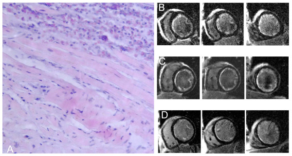
We report the diagnostic potential of cardiovascular magnetic resonance (CMR) to visualize the time course of eosinophilic myocarditis upon successful treatment. A 50-year-old man was admitted with a progressive heart failure. Endomyocardial biopsies were taken from the left ventricle because of a white blood cell count of 17000/mm3 with 41% eosinophils. Histological evaluation revealed endomyocardial eosinophilic infiltration and areas of myocyte necrosis. The patient was diagnosed with hypereosinophilic myocarditis due to idiopathic hypereosinophilic syndrome. CMR-studies at presentation and a follow-up study 3 weeks later showed diffuse subendocardial LGE in the whole left ventricle. Upon treatment with steroids, CMR-studies revealed marked reduction of subendocardial LGE after 3 months in parallel with further clinical improvement. This case therefore highlights the clinical importance of CMR to visualize the extent of endomyocardial involvement in the diagnosis and treatment of eosinophilic myocarditis.
Figures
References
-
- Löffler W. Endocarditis parietalis fibroplastica mit Bluteosinophilie. Schweiz Med Wochenschr. 1936;65:817–820. - PubMed
Publication types
MeSH terms
Substances
LinkOut - more resources
Full Text Sources
Medical
