. 2008;10(3):R41.
doi: 10.1186/bcr2092. Epub 2008 May 8.

Breast cancer tumor growth estimated through mammography screening data

Affiliations

Breast cancer tumor growth estimated through mammography screening data

Harald Weedon-Fekjaer et al. Breast Cancer Res. 2008.

Abstract

Introduction: Knowledge of tumor growth is important in the planning and evaluation of screening programs, clinical trials, and epidemiological studies. Studies of tumor growth rates in humans are usually based on small and selected samples. In the present study based on the Norwegian Breast Cancer Screening Program, tumor growth was estimated from a large population using a new estimating procedure/model.

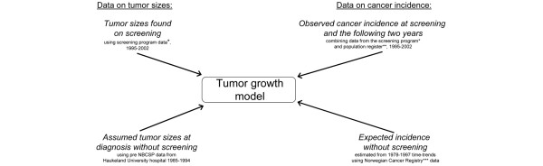
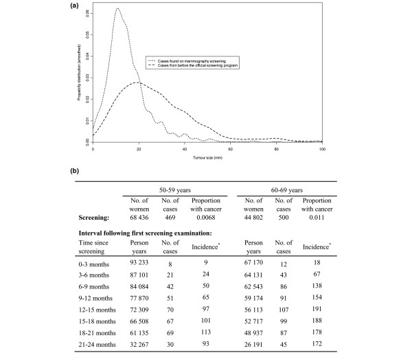
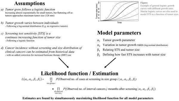
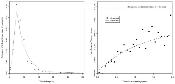
Methods: A likelihood-based estimating procedure was used, where both tumor growth and the screen test sensitivity were modeled as continuously increasing functions of tumor size. The method was applied to cancer incidence and tumor measurement data from 395,188 women aged 50 to 69 years.

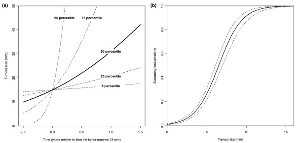
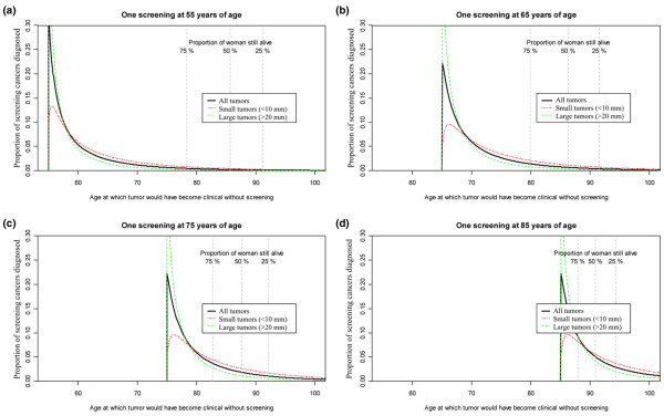
Results: Tumor growth varied considerably between subjects, with 5% of tumors taking less than 1.2 months to grow from 10 mm to 20 mm in diameter, and another 5% taking more than 6.3 years. The mean time a tumor needed to grow from 10 mm to 20 mm in diameter was estimated as 1.7 years, increasing with age. The screen test sensitivity was estimated to increase sharply with tumor size, rising from 26% at 5 mm to 91% at 10 mm. Compared with previously used Markov models for tumor progression, the applied model gave considerably higher model fit (85% increased predictive power) and provided estimates directly linked to tumor size.

Conclusion: Screening data with tumor measurements can provide population-based estimates of tumor growth and screen test sensitivity directly linked to tumor size. There is a large variation in breast cancer tumor growth, with faster growth among younger women.


Figures

Figure 1
Figure 1
Data sources used in the estimation. *Norwegian Breast Cancer Screening Program (NBCSP). **Statistics Norway (SSB). ***Norwegian Cancer Registry.
Figure 2
Figure 2
Summary of data. (a) Distribution of tumor sizes. (b) Observed number of cases at first screening and in the following interval. Tumor measurements from before the official screening program started come from a database at Haukeland Hospital (1985 to 1994). Screening data include only the first appearance of women reporting no earlier screening history, while interval data are based on all available observations. *Cases per 100,000 person-years.
Figure 3
Figure 3
Overview of the new cancer growth model. New cancer growth model: assumptions, model parameters, and likelihood function.
Figure 4
Figure 4
Estimates of tumor growth rate variation and screening test sensitivity for all age groups combined. Estimates for all age groups combined, with correction of background incidence (+21%) due to increased hormone therapy use. (a) Estimated variation of tumor growth rates, illustrated by growth curves for the 5th, 25th, 50th, 75th and 95th percentiles. (b) Estimated screening test sensitivity with 95% pointwise confidence intervals.
Figure 5
Figure 5
Model fit using the new cancer growth model. (Left) Tumor sizes on screening. (Right) Number of interval cancers. HRT = hormone replacement therapy.
Figure 6
Figure 6
Illustration of potential use of the new cancer growth model. Age at which screening tumors would have become clinical without screening, by tumor size at the time of screening detection. (a) Screening at 55 years of age. (b) Screening at 65 years of age. (c) Screening at 75 years of age. (d) Screening at 85 years of age. Vertical lines mark the expected time at which 25%, 50% and 75% of the screened women are suspected to have died, based on death rates from Statistics Norway. Panel (c) and (d) are based on the screening test sensitivity and growth estimates from the 60 to 69 years age group.

References

    1. Berry DA, Cronin KA, Plevritis SK, Fryback DG, Clarke L, Zelen M, Mandelblatt JS, Yakovlev AY, Habbema JD, Feuer EJ. Effect of screening and adjuvant therapy on mortality from breast cancer. N Engl J Med. 2005;353:1784–1792. doi: 10.1056/NEJMoa050518. - DOI - PubMed
    1. Spratt JS, Meyer JS, Spratt JA. Rates of growth of human solid neoplasms: part I. J Surg Oncol. 1995;60:137–146. doi: 10.1002/jso.2930600216. - DOI - PubMed
    1. Spratt JS, Meyer JS, Spratt JA. Rates of growth of human neoplasms: part II. J Surg Oncol. 1996;61:68–83. doi: 10.1002/1096-9098(199601)61:1<68::AID-JSO2930610102>3.0.CO;2-E. - DOI - PubMed
    1. Peer PG, van Dijck JA, Hendriks JH, Holland R, Verbeek AL. Age-dependent growth rate of primary breast cancer. Cancer. 1993;71:3547–3551. doi: 10.1002/1097-0142(19930601)71:11<3547::AID-CNCR2820711114>3.0.CO;2-C. - DOI - PubMed
    1. Prevost TC, Launoy G, Duffy SW, Chen HH. Estimating sensitivity and sojourn time in screening for colorectal cancer: a comparison of statistical approaches. Am J Epidemiol. 1998;148:609–619. - PubMed

Publication types