. 2008 May 8;58(3):333-9.
doi: 10.1016/j.neuron.2008.02.028.

Prestin-based outer hair cell motility is necessary for mammalian cochlear amplification

Affiliations

Prestin-based outer hair cell motility is necessary for mammalian cochlear amplification

Peter Dallos et al. Neuron. .

Abstract

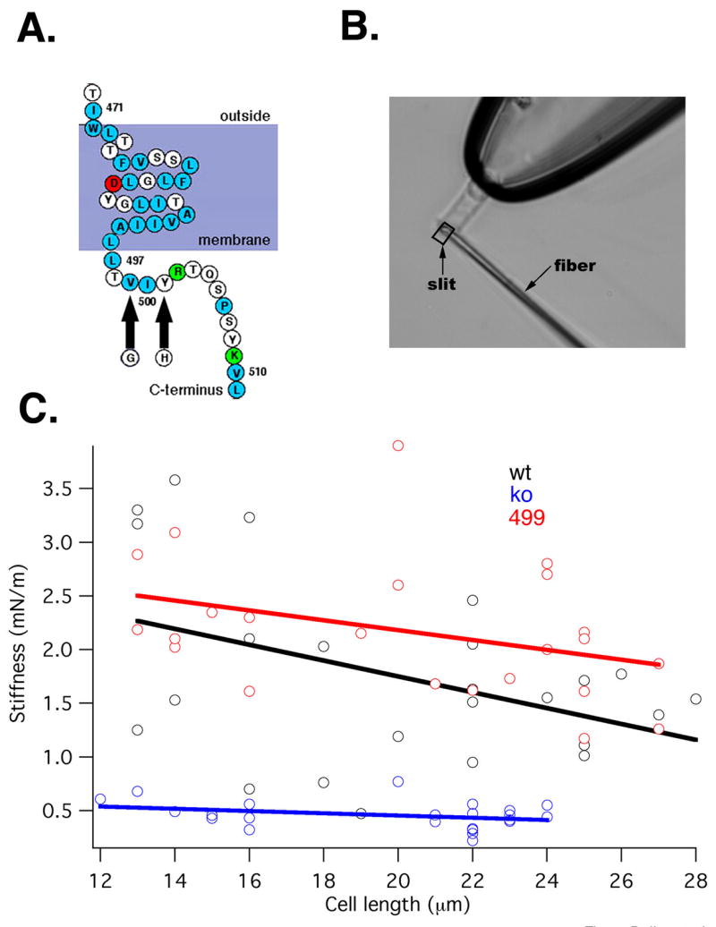
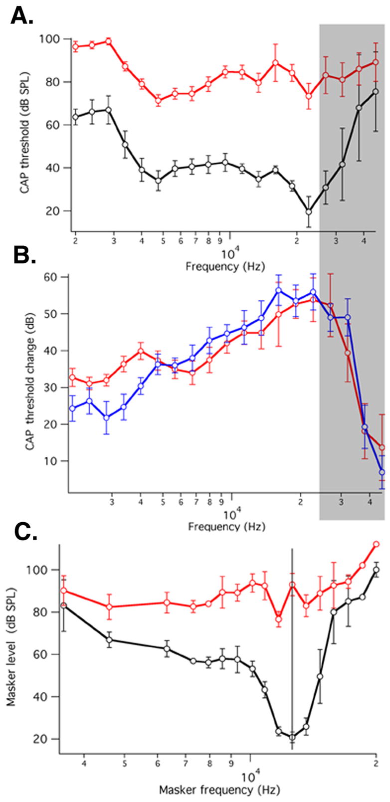
It is a central tenet of cochlear neurobiology that mammalian ears rely on a local, mechanical amplification process for their high sensitivity and sharp frequency selectivity. While it is generally agreed that outer hair cells provide the amplification, two mechanisms have been proposed: stereociliary motility and somatic motility. The latter is driven by the motor protein prestin. Electrophysiological phenotyping of a prestin knockout mouse intimated that somatic motility is the amplifier. However, outer hair cells of knockout mice have significantly altered mechanical properties, making this mouse model unsatisfactory. Here, we study a mouse model without alteration to outer hair cell and organ of Corti mechanics or to mechanoelectric transduction, but with diminished prestin function. These animals have knockout-like behavior, demonstrating that prestin-based electromotility is required for cochlear amplification.


Figures

Figure 1
Figure 1
A. Partial amino-acid sequence of the predicted topology of the prestin molecule showing the last putative membrane-spanning helix and part of the C-terminus. The 499/501 mutation is indicated by arrows. 1.B. Image of a KO mouse OHC held by a suction pipette, along with a driven fiber positioned against the cell’s ciliated pole for the purpose of stiffness determination. The combined displacement of fiber and cell is monitored by a photodiode through a rectangular slit. 1.C. Plots of OHC somatic stiffness vs. cell length. Wild-type (black N=22), prestin KO (blue N=21) and 499 KI (red N=23; this color scheme will be followed in all subsequent plots) are shown. Also presented are linear regression lines fitted to the data.
Figure 2
Figure 2
A. Plasma membrane targeting is examined by immunostaining in OHCs of WT and 499 prestin mice using confocal microscopy. Whole-mount preparations of the apical cochlear turns of WT and 499 mice are shown at P25. Immunofluorescent images derived from WT (first two images) and 499 homozygous mice (second two images) stained with anti-C-mPres. First and third images: treatment with triton X-100, second and fourth images: no treatment. Similar staining patterns were also observed using anti-N-mPres. 2.B. Prestin’s oligomeric status in WT and 499 cochleae examined by NEXT-PAGE/Western blot. EDT= ethanedithiol. 2.C. Intensities of monomer and dimer bands are compared between WT and 499 cochleae. There is no statistical difference between the amounts of monomer (t-test, p=0.11), dimer (p=0.99) or their ratio (p=0.59; N=3).
Figure 3
Figure 3. In vitro measurements of hair cell function
3.A. Average NLC (±S.D) for WT and 499 OHCs (499 NLC N=8; WT NLC N=11). 3.B. Average motility (cell-length change) ±S.D. (WT, N=11; 499, N=9). Cell contraction is plotted up. Linear capacitance values, measured as the asymptotes of the NLC curves at large positive membrane potential for WT and large negative voltage for 499 mice, are similar. (Clin±SD for WT: 7.8±0.9 pF and 499: 7.80±0.66 pF and t-test: p=0.72). Thus, cell-membrane dimensions in the two genotypes, reflected in linear capacitance, are comparable. 3.C. Representative transducer currents in response to 100 Hz sinusoidal displacements of the basilar membrane from one WT and one 499 OHC in the hemicochlea. Amplitude of BM displacement varied between approximately ±120 nm in equal steps. Inward current is plotted down, holding potential is −70 mV. 3.D. Average (±S.D., N=5 WT; N=5 KI) change in transducer current magnitude in response to sinusoidal displacements of the BM. Inward current is plotted down, holding potential is −70 mV. 3.E. Representative transducer currents in response to 20 msec DC displacement of the TM toward scala tympani for one apical WT OHC and a corresponding 499 cell.
Figure 4
Figure 4. In vivo results showing CAP data obtained from a round-window electrode
4.A. Average (±S.D. N=6 WT; N=7 KI) CAP thresholds (sound pressure level required to measure10 μV CAP) as a function of stimulus frequency for 499 KI and WT mice. 4.B. Average differences (±S.D) between individual 499 KI thresholds and the corresponding average WT threshold (red). Average differences (±S.D) between individual prestin KO thresholds and the corresponding average WT threshold (blue). The KO mice used for comparison were described in Cheatham et al. (2007). Shaded region approximately corresponds to extent of age-related hair-cell loss. 4.C. Average (±S.D) CAP masking tuning curves for 499 KI (N=7) and WT mice (N=5). Probe tone frequency is 12 kHz. Inasmuch as our tuning-curve collection platform uses slightly different masker frequencies for each tuning curve, an average frequency scale was created. As a consequence, the average WT tuning curve appears to be more shallowly tuned than those published previously (Dallos and Cheatham, 1976; Cheatham et al., 2004; 2007).

Comment in

References

    1. Ashmore JF. Forward and reverse transduction in the mammalian cochlea. Neurosci Res Suppl. 1990;12:S39–S50. - PubMed
    1. Brownell W, Bader CR, Bertrand D, de Ribaupierre Y. Evoked mechanical responses of isolated cochlear outer hair cells. Science. 1985;227:194–196. - PubMed
    1. Chan D, Hudspeth AJ. Ca2+ current-driven nonlinear amplification by the mammalian cochlea in vitro. Nat Neurosci. 2005;8:149–155. - PMC - PubMed
    1. Cheatham MA, Dallos P. Longitudinal comparisons of IHC AC and DC receptor potentials recorded from the guinea-pig cochlea. Hear Res. 1993;68:107–114. - PubMed
    1. Cheatham MA, Huynh KH, Gao J, Zuo J, Dallos P. Cochlear function in Prestin knockout mice. J Physiol (London) 2004;560:821–830. - PMC - PubMed

Publication types

Substances

LinkOut - more resources