. 2008 Jun;162(3):404-10.
doi: 10.1016/j.jsb.2008.02.010. Epub 2008 Mar 31.

Mechanical properties of mineralized collagen fibrils as influenced by demineralization

Affiliations

Mechanical properties of mineralized collagen fibrils as influenced by demineralization

M Balooch et al. J Struct Biol. 2008 Jun.

Abstract

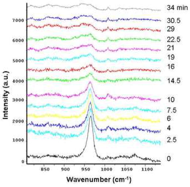
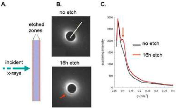
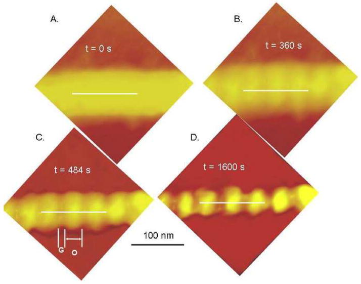
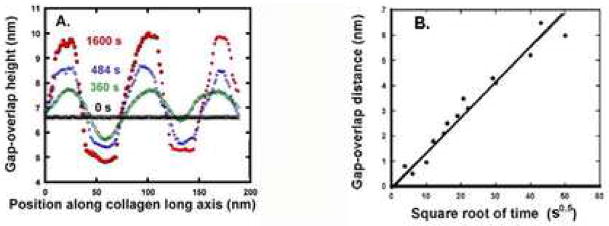
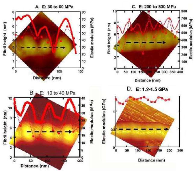
Dentin and bone derive their mechanical properties from a complex arrangement of collagen type-I fibrils reinforced with nanocrystalline apatite mineral in extra- and intrafibrillar compartments. While mechanical properties have been determined for the bulk of the mineralized tissue, information on the mechanics of the individual fibril is limited. Here, atomic force microscopy was used on individual collagen fibrils to study structural and mechanical changes during acid etching. The characteristic 67 nm periodicity of gap zones was not observed on the mineralized fibril, but became apparent and increasingly pronounced with continuous demineralization. AFM-nanoindentation showed a decrease in modulus from 1.5 GPa to 50 MPa during acid etching of individual collagen fibrils and revealed that the modulus profile followed the axial periodicity. The nanomechanical data, Raman spectroscopy and SAXS support the hypothesis that intrafibrillar mineral etches at a substantially slower rate than the extrafibrillar mineral. These findings are relevant for understanding the biomechanics and design principles of calcified tissues derived from collagen matrices.


Figures

Figure 1
Figure 1
Raman spectra during demineralization of 50 mg powder specimens of human dentin. The phosphate peak at 960 cm−1 decreased ~ 80% after 10 min. and was not distinguishable from background at 29 min. A small peak at 1007 cm−1 attributed to phyenylanine increased slightly in the first 10 min. and remained essentially constant subsequently.
Figure 2
Figure 2
SAXS experiment demonstrating enhancement of the 67 nm (22 nm third harmonic, red arrows) associated with intrafibrillar mineral in collagen fibrils. A. experimental set up showing x-ray beam penetrating the dentin slab. B. SAXS patterns were obtained after the beam penetrated the unetched dentin slab (top) and after etching (bottom) for 16 h in 10% citric acid so that the beam penetrated two surfaces that had been demineralized (etch zones in A.) and an unetched central core. White line in the top pattern shows the line used for the intensity profile. C. Intensity profiles before (black line) and after (red line) the 16 h etching experiment. Red arrows in B and C show the development of a pronounced peak associated with the third harmonic of scattering from the intrafibrillar mineral with 67 nm periodicity from apatite deposited in the gap zones of mineralized collagen fibrils (Kinney et al, 2001a). The SAXS profiles were adjusted so that the 16 h etch had the same maximum value as the unetched data. This normalization was helpful in order to show the difference between the two scattering curves in the same q range for scattering.
Figure 3
Figure 3
Demineralization sequence in 10% citric acid of an isolated human dentin collagen fibril with white lines showing lines used to measure height variations in Fig. 4. A. initial appearance of the fibril was smooth and appeared to have indistinct boundaries. B. After 360 s of demineralization the underlying periodicity of the collagen fibril due to gap-overlap height differences was apparent. C. gap-overlap height differences increased with further demineralization at 484 s, labels G and O indicate gap and overlap zones, respectively. D.1600 s demineralization..
Figure 4
Figure 4
A. Gap-overlap height differences of the collagen fibril shown in Fig. 3 (along white lines). The flat topography of the fibril (black line) increased with progressive demineralization shown at 360 s (green), 484 s (blue) and 1600 s (red). Similar results were seen in other fibrils demineralized for up to 3600 s. B. Gap-overlap distance increases with demineralization time for several human dentin collagen fibrils plotted vs. square root of time (s), suggesting a slow diffusion controlled process.
Figure 5
Figure 5
AFM images associating topographic variations (solid red lines) with indentation elastic modulus values (broken red lines) along black dashed lines on isolated collagen fibrils with varying appearance. A. recombinant collagen fibril without mineral had gap-overlap height differences of 5 nm. Modulus values varied from about 30 MPa at gaps to 60 MPa at the overlaps. B. Fully demineralized human dentin collagen fibrils showing gap overlap height differences of about 6 nm and modulus variations between 10 MPa at gaps to 40 MPa in the overlap zones. C. Dentin collagen fibril after 240 s demineralization showing sinusoidal periodic variations in topography with gap-overlap height differences of 2–3 nm. Modulus values at the gaps were 200–400 MPa and at the overlaps were 500–800 MPa. D. Smooth dentin collagen fibril showing minimal gap-overlap height differences and modulus variations between 1.2 and 1.5 GPa.

References

    1. Angker L, Nijhof N, Swain MV, Kilpatrick NM. Influence of hydration and mechanical characterization of carious primary dentine using an ultra-micro indentation system (UMIS) Eur J Oral Sci. 2004;112:231–236. - PubMed
    1. Angker L, Swain MV, Kilpatrick N. Characterizing the micro-mechanical behavior of the carious dentine of primary teeth using nano-indentation. J Biomech. 2005;38:1535–1542. - PubMed
    1. Balooch M, Wu-Magidi IC, Lindquist AS, Balazs, Marshall SJ, Marshall GW, Seikhaus WJ, Kinney JH. Viscolelastic Properties of Demineralized Human Dentin in Water with AFM-Based Indentation. J Biomed Mater Res. 1998;40 :539–544. - PubMed
    1. Bonar LC, Lee S, Mook HA. Neutron diffraction studies of collagen in fully mineralized bone. J Molec Biol. 1985;181:265–270. - PubMed
    1. Carrillo F, Gupta S, Balooch M, Marshall SJ, Marshall GW, Pruitt L, Puttlitz C. Nanoindentation of polydimethylsiloxane elastomers: Effect of crosslinking, work of adhesion and fluid environment on elastic modulus. J Mater Res. 2005:2820–2830.

Publication types

LinkOut - more resources