. 2008 Aug;149(8):3799-808.
doi: 10.1210/en.2008-0117. Epub 2008 May 8.

Epidermal growth factor induces G protein-coupled receptor 30 expression in estrogen receptor-negative breast cancer cells

Affiliations

Epidermal growth factor induces G protein-coupled receptor 30 expression in estrogen receptor-negative breast cancer cells

Lidia Albanito et al. Endocrinology. 2008 Aug.

Abstract

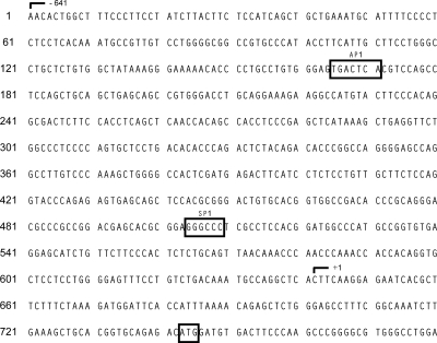
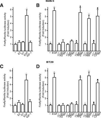
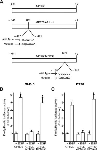
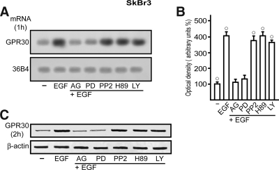
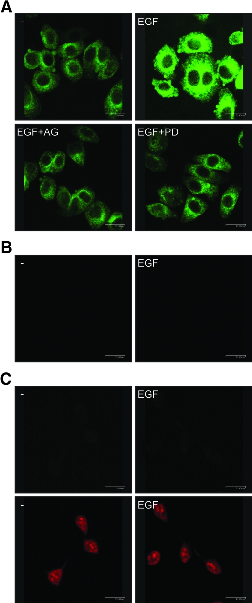
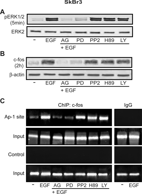
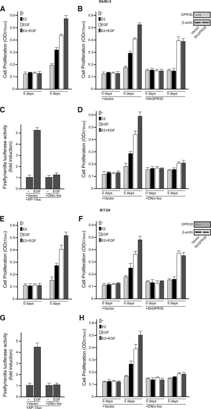
Different cellular receptors mediate the biological effects induced by estrogens. In addition to the classical nuclear estrogen receptors (ERs)-alpha and -beta, estrogen also signals through the seven-transmembrane G-protein-coupled receptor (GPR)-30. Using as a model system SkBr3 and BT20 breast cancer cells lacking the classical ER, the regulation of GPR30 expression by 17beta-estradiol, the selective GPR30 ligand G-1, IGF-I, and epidermal growth factor (EGF) was evaluated. Transient transfections with an expression plasmid encoding a short 5'-flanking sequence of the GPR30 gene revealed that an activator protein-1 site located within this region is required for the activating potential exhibited only by EGF. Accordingly, EGF up-regulated GPR30 protein levels, which accumulated predominantly in the intracellular compartment. The stimulatory role elicited by EGF on GPR30 expression was triggered through rapid ERK phosphorylation and c-fos induction, which was strongly recruited to the activator protein-1 site found in the short 5'-flanking sequence of the GPR30 gene. Of note, EGF activating the EGF receptor-MAPK transduction pathway stimulated a regulatory loop that subsequently engaged estrogen through GPR30 to boost the proliferation of SkBr3 and BT20 breast tumor cells. The up-regulation of GPR30 by ligand-activated EGF receptor-MAPK signaling provides new insight into the well-known estrogen and EGF cross talk, which, as largely reported, contributes to breast cancer progression. On the basis of our results, the action of EGF may include the up-regulation of GPR30 in facilitating a stimulatory role of estrogen, even in ER-negative breast tumor cells.


Figures

Figure 1
Figure 1
Sequence of the GPR30–5′-flanking region used to generate luciferase reporter constructs.
Figure 2
Figure 2
The GPR30–5′-flanking region is transactivated by EGF in SkBr3 and BT20 breast cancer cells. A–D, Cells were transfected with a reporter plasmid encoding the GPR30–5′-flanking region and treated with 100 nm E2, 1 μm G-1, 50 ng/ml IGF-I, or 50 ng/ml EGF, and 10 μm EGFR inhibitor tyrphostin AG 1478 (AG), 10 μm MEK inhibitor PD, 10 μm Src family tyrosine kinase inhibitor PP2, 10 μm PKA inhibitor H89, 10 μm PI3K inhibitor LY, as indicated. The luciferase activities were normalized to the internal transfection control and values of cells receiving vehicle (−) were set as 1-fold induction upon which the activity induced by treatments was calculated. Each data point represents the mean ± sd of three independent experiments performed in triplicate. ○, •, □, ▪, P < 0.05, for cells receiving vehicle (−) vs. treatment.
Figure 3
Figure 3
An AP1 site is responsible for transactivation of the GPR30–5′-flanking region induced by EGF in SkBr3 and BT20 breast cancer cells. A, AP1 (GPR30AP1mut) and SP1 (GPR30SP1mut) mutations generated within the GPR30–5′-flanking region. B and C, Cells were transfected with the reporter plasmids described in A and treated with 50 ng/ml EGF. The luciferase activities were normalized to the internal transfection control and values of cells receiving vehicle (−) were set as 1-fold induction upon which the activity induced by treatments was calculated. Each data point represents the mean ± sd of three independent experiments performed in triplicate. ○, •, P < 0.05, for cells receiving vehicle (−) vs. treatment.
Figure 4
Figure 4
EGF up-regulates GPR30 expression in SkBr3 cells. A, The expression of GPR30 was evaluated by semiquantitative RT-PCR in cells treated for 1 h with vehicle (−) or 50 ng/ml EGF alone and in combination with 10 μm EGFR inhibitor tyrphostin AG, 10 μm MEK inhibitor PD, 10 μm Src family tyrosine kinase inhibitor PP2, 10 μm PKA inhibitor H89, 10 μm PI3K inhibitor LY, as indicated. The housekeeping gene 36B4 was determined as a control. B, Quantitative representation of GPR30 mRNA expression (mean ± sd) of three independent experiments after densitometry and correction for 36B4 expression. ○, P < 0.05, for cells receiving vehicle (−) vs. treatment. C, Immunoblot of GPR30 from SkBr3 cells treated for 2 h with vehicle (−) or 50 ng/ml EGF alone and in combination with 10 μm AG, 10 μm PD, 10 μm PP2, 10 μm H89, and 10 μm LY, as indicated. β-Actin served as a loading control.
Figure 5
Figure 5
GPR30 localization in SkBr3 cells. A, GPR30 evaluation by confocal microscopy in SkBr3 cells fixed, permeabilized, and stained with anti-GPR30 antibody. Cells were treated for 2 h with vehicle (−) or 50 ng/ml EGF alone and in combination with EGFR inhibitor tyrphostin AG, 10 μm MEK inhibitor PD, as indicated. B, SkBr3 cells were treated for 2 h with vehicle (−) or 50 ng/ml EGF and stained with GPR30 antibody, which was preneutralized with the antigen peptide. C, HEK-293 cells were treated for 2 h with vehicle (−) or 50 ng/ml EGF and stained with GPR30 antibody (upper panels) or propidium iodide (lower panels). The white bars denote 21,43 μm. Data are representative of three independent experiments.
Figure 6
Figure 6
EGF induces ERK1/2 phosphorylation and c-fos expression, which is recruited at the AP1 site located in the GPR30–5′-flanking region. A, The rapid ERK1/2 phosphorylation induced by 50 ng/ml EGF in SkBr3 cells is abrogated in presence of 10 μm EGFR inhibitor tyrphostin AG and 10 μm MEK inhibitor PD but not in presence of 10 μm Src family tyrosine kinase inhibitor PP2, 10 μm PKA inhibitor H89, or 10 μm PI3K inhibitor LY. B, The up-regulation of c-fos induced by 50 ng/ml EGF in SkBr3 cells is abrogated in presence of 10 μm AG and 10 μm PD but not in presence of 10 μm PP2, 10 μm H89, or 10 μm LY. C, EGF treatment (50 ng/ml) induces in SkBr3 cells the recruitment of c-fos at the AP1 site located in the GPR30–5′-flanking region. This recruitment is abrogated by 10 μm AG or 10 μm PD but persists in presence of 10 μm PP2, 10 μm H89, or 10 μm LY. The amplification of a region lacking the AP1 site (control) does not show the recruitment following the same experimental conditions described above. In control samples, nonspecific IgG was used instead of the primary antibody.
Figure 7
Figure 7
In SkBr3 and BT20 breast cancer cells, EGF engages E2 through GPR30 to boost the growth effects, which were monitored by MTT assay. A and E, The combination of E2 and EGF treatments enhances the proliferation of SkBr3 and BT20 cells stimulated by each mitogen used alone. Cells were treated with vehicle or 100 nm E2 and/or 50 ng/ml EGF in medium containing 2.5% charcoal-stripped FBS (medium was refreshed and treatments were renewed every 2 d). B and F, The growth effects induced by E2 alone or in combination with EGF were abolished by GPR30 silencing in both SkBr3 and BT20 cells. Cells were transfected with an empty vector or shGPR30 and the next day were treated with vehicle (−), 100 nm E2, and/or 50 ng/ml EGF. Transfections and treatments were renewed every 2 d. Efficacy of GPR30 silencing was evaluated by immunoblots, as indicated. C and G, The DN/c-fos construct effectively blocked the AP1 mediated transcriptional activity in SkBr3 and BT20 cells. The luciferase activities were normalized to the internal transfection control and values of cells receiving vehicle (−) were set as 1-fold induction upon which the activity induced by 50 ng/ml EGF was calculated. D and H, The growth effects induced by E2 and EGF used alone or in combination were abolished transfecting the SkBr3 and BT20 cells with the DN/c-fos expression vector. Cells were transfected with an empty vector or the DN/c-fos construct and the next day were treated with vehicle (−), 100 nm E2, and/or 50 ng/ml EGF. Each data point is the mean ± sd of three independent experiments performed in triplicate.

Similar articles

Cited by

References

    1. Levin ER 2003 Bidirectional signaling between the estrogen receptor and the epidermal growth factor receptor. Mol Endocrinol 17:309–317 - PubMed
    1. Keen JC, Davidson NE 2003 The biology of breast carcinoma. Cancer 97:825–833 - PubMed
    1. Roskoski Jr R 2004 The ErbB/HER receptor protein-tyrosine kinases and cancer. Biochem Biophys Res Commun 319:1–11 - PubMed
    1. Ali S, Coombes RC 2002 Endocrine-responsive breast cancer and strategies for combating resistance. Nat Rev Cancer 2:101–112 - PubMed
    1. Razandi M, Pedram A, Park ST, Levin ER 2003 Proximal events in signaling by plasma membrane estrogen receptor. J Biol Chem 278:2701–2712 - PubMed

Publication types

MeSH terms