. 2008 Jul;147(3):1072-91.
doi: 10.1104/pp.107.109512. Epub 2008 May 8.

The beta-glucosidases responsible for bioactivation of hydroxynitrile glucosides in Lotus japonicus

Affiliations

The beta-glucosidases responsible for bioactivation of hydroxynitrile glucosides in Lotus japonicus

Anne Vinther Morant et al. Plant Physiol. 2008 Jul.

Erratum in

  • Plant Physiol. 2010 Apr;152(4):2269

Abstract

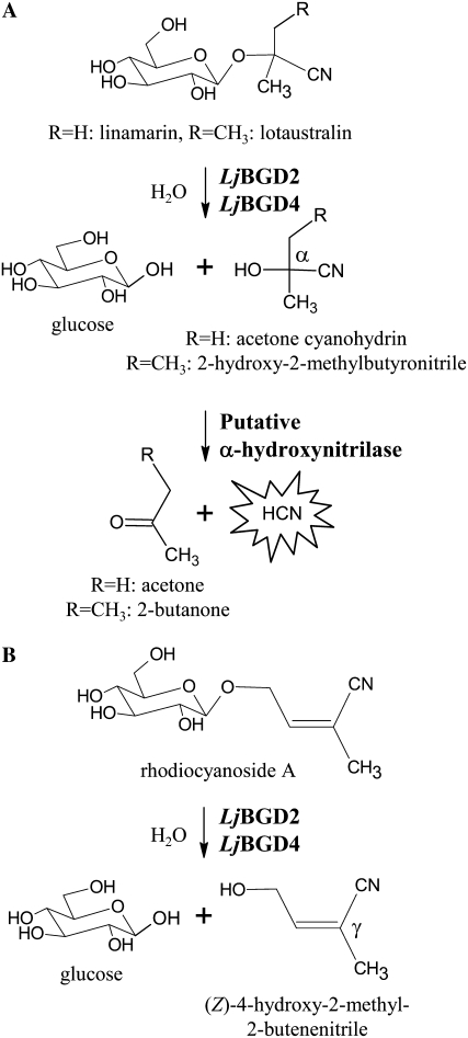
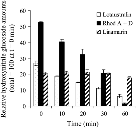
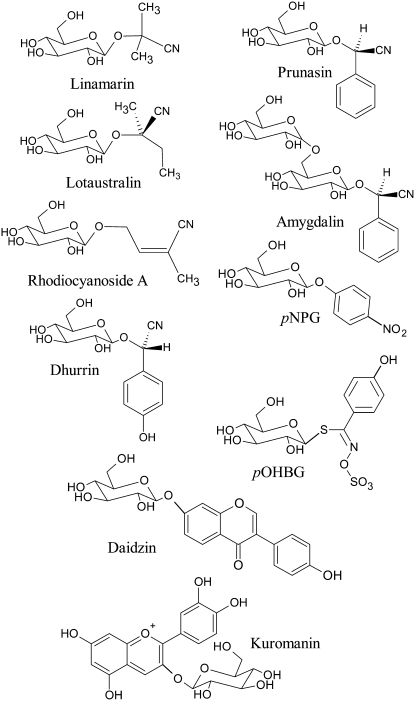
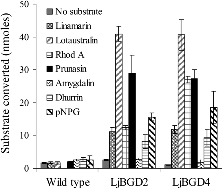
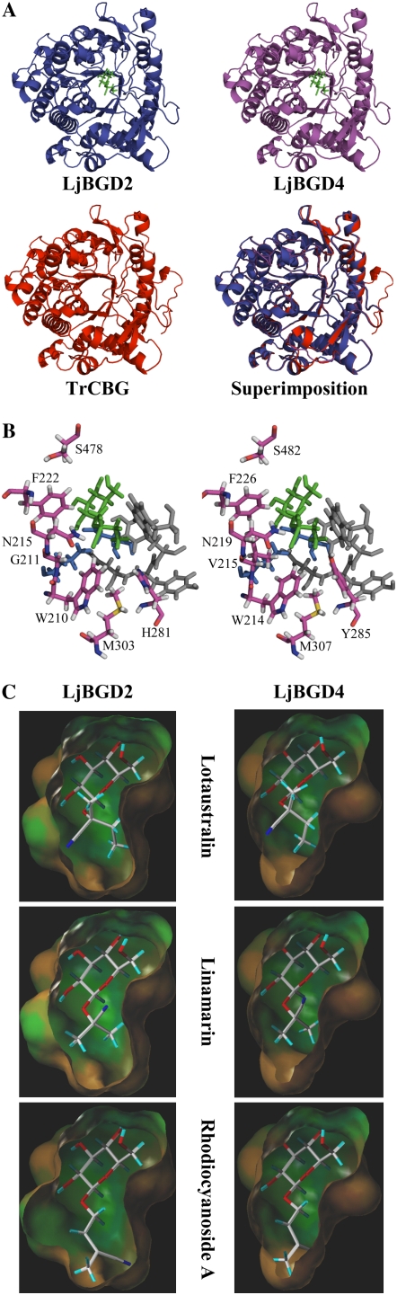
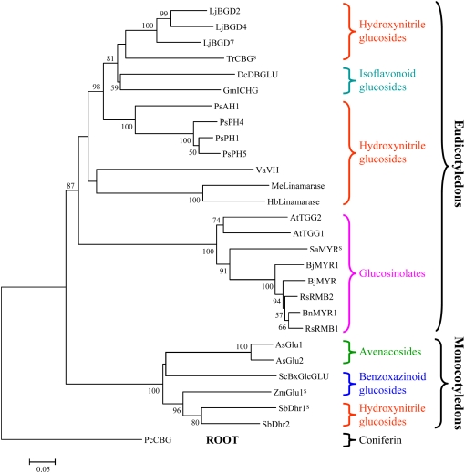
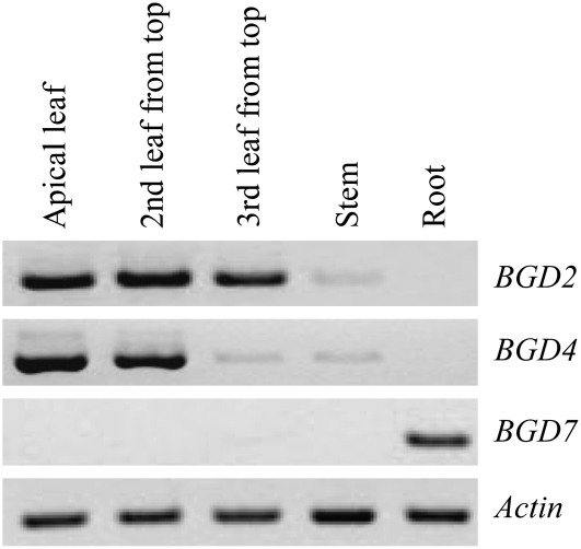
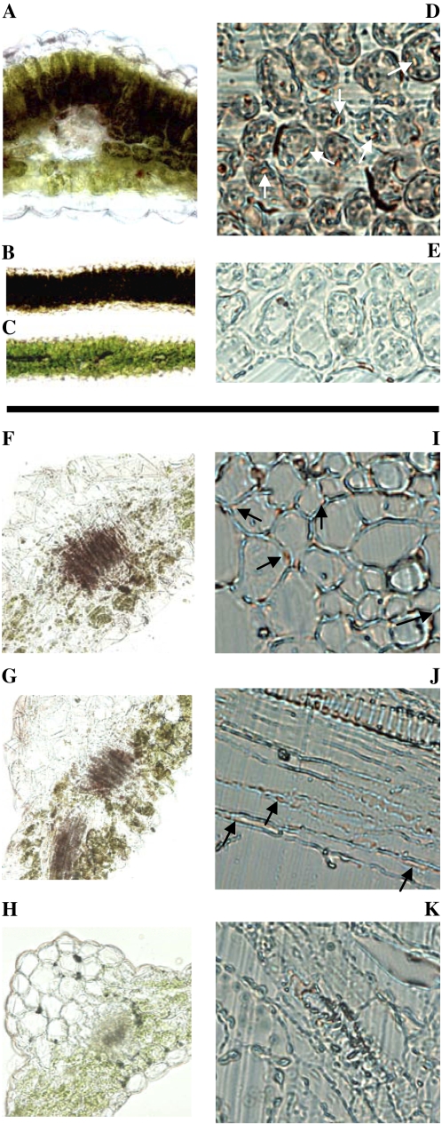
Lotus japonicus accumulates the hydroxynitrile glucosides lotaustralin, linamarin, and rhodiocyanosides A and D. Upon tissue disruption, the hydroxynitrile glucosides are bioactivated by hydrolysis by specific beta-glucosidases. A mixture of two hydroxynitrile glucoside-cleaving beta-glucosidases was isolated from L. japonicus leaves and identified by protein sequencing as LjBGD2 and LjBGD4. The isolated hydroxynitrile glucoside-cleaving beta-glucosidases preferentially hydrolyzed rhodiocyanoside A and lotaustralin, whereas linamarin was only slowly hydrolyzed, in agreement with measurements of their rate of degradation upon tissue disruption in L. japonicus leaves. Comparative homology modeling predicted that LjBGD2 and LjBGD4 had nearly identical overall topologies and substrate-binding pockets. Heterologous expression of LjBGD2 and LjBGD4 in Arabidopsis (Arabidopsis thaliana) enabled analysis of their individual substrate specificity profiles and confirmed that both LjBGD2 and LjBGD4 preferentially hydrolyze the hydroxynitrile glucosides present in L. japonicus. Phylogenetic analyses revealed a third L. japonicus putative hydroxynitrile glucoside-cleaving beta-glucosidase, LjBGD7. Reverse transcription-polymerase chain reaction analysis showed that LjBGD2 and LjBGD4 are expressed in aerial parts of young L. japonicus plants, while LjBGD7 is expressed exclusively in roots. The differential expression pattern of LjBGD2, LjBGD4, and LjBGD7 corresponds to the previously observed expression profile for CYP79D3 and CYP79D4, encoding the two cytochromes P450 that catalyze the first committed step in the biosyntheis of hydroxynitrile glucosides in L. japonicus, with CYP79D3 expression in aerial tissues and CYP79D4 expression in roots.


Figures

Figure 1.
Figure 1.
Bioactivation of lotaustralin, linamarin, and rhodiocyanoside A in L. japonicus leaves. A, Cyanogenic glucosides are β-glucosides of α-hydroxynitriles. Upon cell disruption, β-glucosidases catalyze the hydrolysis of the O-β-glucosidic bond to yield Glc and an α-hydroxynitrile aglucone. The α-hydroxynitrile either spontaneously or enzymatically breaks down to liberate a ketone and toxic HCN. B, Rhodiocyanosides are β-glucosides of β- or γ-hydroxynitriles. In contrast to α-hydroxynitrile glucosides, aglucone formation is not accompanied by the release of HCN.
Figure 2.
Figure 2.
Degradation of lotaustralin, linamarin, and rhodiocyanosides in leaves of L. japonicus following cell disruption. The rate of degradation of endogenous hydroxynitrile glucosides in transgenic L. japonicus 35S∷CYP79D2 leaves following tissue disruption was monitored by LC-MS and analysis of the amounts of metabolites remaining at time points between 0 and 60 min following cell disruption. The total amount of hydroxynitrile glucosides (linamarin + lotaustralin + rhodiocyanosides) at time 0 was defined as 100. Rhodiocyanosides are most rapidly degraded, followed by lotaustralin, which is degraded at a moderate rate; only negligible degradation of linamarin is observed.
Figure 3.
Figure 3.
Purification of hydroxynitrile glucoside-cleaving β-glucosidases from L. japonicus leaves. Protein fractions obtained during the purification procedure were analyzed by 12% SDS-PAGE, and the protein composition was visualized by Coomassie Blue staining. The lanes show protein composition in the total crude leaf extract (1), soluble crude leaf extract (2), cation exchange supernatant (3), cation exchange eluate (4), and fractions from gel filtration chromatography containing β-glucosidase activity against hydroxynitrile glucosides (5). The presence (+) or absence (÷) of β-glucosidase activity toward lotaustralin and linamarin as measured by HCN release is indicated at the top of each lane. Protein sequencing identified the protein bands indicated by A and B as a mixture of LjBGD2 and LjBGD4, while the faster migrating protein band was identified as Rubisco.
Figure 4.
Figure 4.
Structures of the main β-glucosides used to examine the substrate specificity of the hydroxynitrile glucoside-cleaving β-glucosidases from L. japonicus. Val-derived linamarin and Ile-derived lotaustralin and rhodiocyanoside A are synthesized by L. japonicus. The α-hydroxynitrile glucosides dhurrin and prunasin are derived from Tyr and Phe, respectively. Amygdalin is the diglucoside derived from glucosylation of prunasin. pNPG is an artificial chromogenic β-glucoside degraded by a wide range of β-glycosidases. Daidzin and kuromanin are isoflavonoid and flavonoid glucosides, respectively. All glucosides tested contain an O-β-glucosidic bond except for the glucosinolates, which contain an S-β-glucosidic bond.
Figure 5.
Figure 5.
Substrate specificity profiles of LjBGD2 and LjBGD4 expressed in Arabidopsis. Arabidopsis leaf discs producing recombinant LjBGD2 and LjBGD4 were assayed for the ability to hydrolyze a range of β-glycosides. Rhod A, Rhodiocyanoside A. All incubation assays (10 min) were conducted using 1 mm substrate corresponding to a total of 200 nmol. Arabidopsis wild-type leaf discs served as a negative control.
Figure 6.
Figure 6.
Models of the three-dimensional architecture of the substrate-binding pockets of LjBGD2 and LjBGD4 with docking of lotaustralin, linamarin, and rhodiocyanoside A. A, Comparison of the backbone configurations of the modeled LjBGD2 and LjBGD4 monomers and the known structure of TrCBG. A superimposition of the three structures illustrates the highly conserved structure of α-hydroxynitrile glucoside-cleaving β-glucosidases. B, Stick models of the amino acids lining the aglucone-binding pockets of LjBGD2 and LjBGD4. Lotaustralin (green) is docked with the two catalytic Glu residues (E208 and E420 [LjBGD2 numbering], shown in blue) positioned on both sides of the β-glucosidic bond. Four highly conserved amino acids (N349, Y350, Y351, and W392 [LjBGD2 numbering], shaded in gray) line one side of the aglucone-binding pocket, while the amino acids lining the opposite side (magenta) are highly variable. Only the unconserved amino acids are specified. The LjBGD2 and LjBGD4 aglucone-binding pockets differ at two positions (G211/V215 and H281/Y285). The amino acid numbering corresponds to that applied in Supplemental Figure S1. The amino acids that define the glucone-binding sites are highly conserved in all β-glucosidases belonging to the family 1 glycoside hydrolases and are not shown for reasons of simplicity. C, Lipophilic surface representations of the active sites of LjBGD2 and LjBGD4. Lipophilic areas are shown in brown, hydrophilic areas in blue, and neutral areas in green. The orientation of the active site is the same as in B. The three endogenous L. japonicus substrates lotaustralin, linamarin, and rhodiocyanoside A are docked into the active sites.
Figure 7.
Figure 7.
Phylogenetic analysis of selected plant β-glucosidases involved in the bioactivation of defense compounds. The phylogenetic tree includes hydroxynitrile and isoflavonoid glucoside-cleaving β-glucosidases from eudicotyledons, glucosinolate-degrading myrosinases (Brassicales), and selected β-glucosidases involved in the bioactivation of defense compounds in monocotyledons. The defense compounds degraded are indicated for the different groups of β-glucosidases. “S” indicates enzymes for which the crystal structures have been solved. LjBGD2, LjBGD4, and LjBGD7, L. japonicus β-glucosidases (this study); TrCBG, T. repens cyanogenic β-glucosidase (Barrett et al., 1995); GmICHG, G. max isoflavone conjugate-hydrolyzing β-glucosidase (Suzuki et al., 2006); DcBDGLU, D. cochinchinensis dalcochinase (Cairns et al., 2000); PsAH1, PsPH1, PsPH4, and PsPH5, P. serotina amygdalin hydrolase and prunasin hydrolase isoenzymes (Kuroki and Poulton, 1987; Zheng and Poulton, 1995; Zhou et al., 2002); VaVH, Vicia angustifolia vicianin hydrolase (Ahn et al., 2007); MeLinamarase, M. esculenta linamarase (Hughes et al., 1992; Keresztessy et al., 2001); HbLinamarase, H. brasiliensis linamarase (Selmar et al., 1987); AtTGG1 and AtTGG2, Arabidopsis myrosinases (Barth and Jander, 2006); SaMYR, Sinapis alba (white mustard) myrosinase (Burmeister et al., 1997); BjMYR and BjMYR1, Brassica juncea (mustard greens) myrosinases (Heiss et al., 1999); RsRMB1 and RsRMB2, Raphanus sativus (radish) myrosinases (Hara et al., 2000); BnMYR1, Brassica napus (rape) myrosinase (Chen and Halkier, 1999); As-Glu-1 and As-Glu-2, A. sativa avenacosidases (Gusmayer et al., 1994; Kim et al., 2000); ScBxGlcGLU, S. cereale DIBOA-Glc β-glucosidase (Nikus et al., 2003); Zm-Glu-1, Z. mays glucosidase 1 (Czjzek et al., 2001); SbDhr1 and SbDhr2, S. bicolor dhurrinases (Hösel et al., 1987; Verdoucq et al., 2004); PcCBG, Pinus contorta (lodgepole pine) coniferin β-glucosidase (Dharmawardhana et al., 1995, 1999). The bootstrapped neighbor-joining tree was built in MEGA 4.0 (Tamura et al., 2007). The tree was bootstrapped with 1,000 iterations (node cutoff value, 50%). The underlying amino acid sequences in fastA format and the multiple alignment can be accessed at http://www.p450.kvl.dk/VintherMorant_etal_Figure7.tfa and http://www.p450.kvl.dk/VintherMorant_etal_Figure7_Alignment.pdf, respectively. The phylogenetic tree was rooted using PcCBG as an outgroup. For the bootstrap analysis, 1,000 trials were performed, and the bootstrap values are shown in percentages; bootstrap node values below 50% are not shown. A more elaborate phylogenetic analysis of plant β-glycosidases, including those presented here, is available at http://www.p450.kvl.dk/BGD.shtml.
Figure 8.
Figure 8.
Expression profiles of LjBGD2, LjBGD4, and LjBGD7 in 21-d-old L. japonicus seedlings. cDNA produced from mRNA isolated from different tissues was obtained from Forslund et al. (2004), and PCR was performed with specific primers amplifying LjBGD2, LjBGD4, LjBGD7, and ACTIN. LjBGD2 and LjBGD4 are detected in aerial tissues, while LjBGD7 is detected exclusively in roots.
Figure 9.
Figure 9.
Localization of β-glucosidase activity in leaves of L. japonicus wild type (A–E) and transgenic Arabidopsis expressing LjBGD2 and LjBGD4 (F–K). β-Glucosidase activity is represented by red/brown staining resulting from the hydrolysis of the artificial substrate BNG and subsequent aglucone complex formation with Fast Blue BB salt. Cross sections (80 μm) of apical leaves (fresh tissue) of L. japonicus wild type show strong color development in leaf palisade tissue in the presence of BNG and Fast Blue BB salt (A and B), while no staining is observed in the absence of BNG (C). A represents a 20× magnification, while B and C show 5× magnifications. Visualization of the subcellular localization of β-glucosidase activity is possible at 40× magnification of 6-μm cross sections of fixed apical leaf tissue. Application of BNG results in strong staining (indicated by white arrows) localized to the symplast (D), whereas only diffuse and weak apoplastic background color development is observed in the absence of BNG (E). BNG staining for β-glucosidase activity in 80-μm cross sections (20× magnification, fresh tissue) of rosette leaves of Arabidopsis expressing LjBGD2 (F), LjBGD4 (G), and the wild type (H) show that LjBGD2 and LjBGD4 both hydrolyze BNG but no BNG hydrolysis is observed in Arabidopsis wild type. I, J, and K are 40× magnifications of 10-μm sections of fixed Arabidopsis rosette leaves. I shows the subcellular localization of LjBGD2 in a cross section (black arrows). J illustrates the subcellular localization of LjBGD4 in a longitudinal section (black arrows). Very weak apoplastic background staining is observed around the xylem in Arabidopsis wild type (K) as well as in transgenic Arabidopsis. Identical subcellular and tissue localizations of LjBGD2 and LjBGD4 activity are observed upon heterologous expression of the enzymes in Arabidopsis, with activity localized in the symplast at the subcellular level (in agreement with L. japonicus) and in and around the vascular tissue at the tissue level (in contrast to L. japonicus).

References

    1. Ahn YO, Saino H, Mizutani M, Shimizu B, Sakata K (2007) Vicianin hydrolase is a novel cyanogenic β-glycosidase specific to β-vicianoside (6-O-α-L-arabinopyranosyl-β-D-glucopyranoside) in seeds of Vicia angustifolia. Plant Cell Physiol 48 938–947 - PubMed
    1. Bak S, Feyereisen R (2001) The involvement of two P450 enzymes, CYP83B1 and CYP83A1, in auxin homeostasis and glucosinolate biosynthesis. Plant Physiol 127 108–118 - PMC - PubMed
    1. Bak S, Kahn RA, Nielsen HL, Møller BL, Halkier BA (1998) Cloning of three A-type cytochromes P450, CYP71E1, CYP98, and CYP99 from Sorghum bicolor (L.) Moench by a PCR approach and identification by expression in Escherichia coli of CYP71E1 as a multifunctional cytochrome P450 in the biosynthesis of the cyanogenic glucoside dhurrin. Plant Mol Biol 36 393–405 - PubMed
    1. Bak S, Olsen CE, Petersen BL, Møller BL, Halkier BA (1999) Metabolic engineering of p-hydroxybenzylglucosinolate in Arabidopsis by expression of the cyanogenic CYP79A1 from Sorghum bicolor. Plant J 20 663–671 - PubMed
    1. Bak S, Paquette S, Morant M, Morant AV, Saito S, Bjarnholt N, Zagrobelny M, Jørgensen K, Osmani S, Hamann T, et al (2006) Cyanogenic glucosides: a case study for evolution and application of cytochromes P450. Phytochem Rev 5 309–329

Publication types

MeSH terms

LinkOut - more resources