Review
. 2008 Sep;84(3):623-30.
doi: 10.1189/jlb.1107762. Epub 2008 May 8.

Macrophages define the invasive microenvironment in breast cancer

Affiliations
Review

Macrophages define the invasive microenvironment in breast cancer

Jeffrey W Pollard. J Leukoc Biol. 2008 Sep.

Abstract

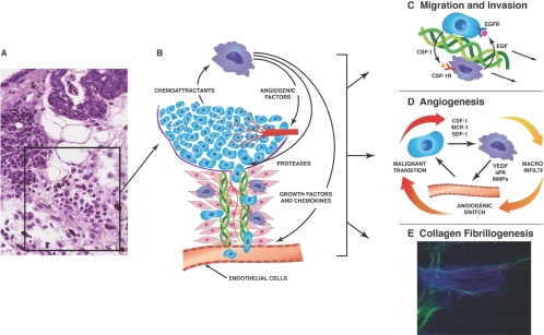
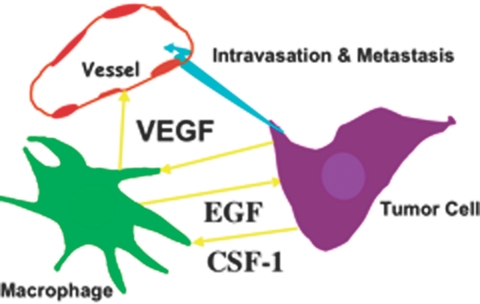
In many human cancers, the abundance of macrophages in the tumor microenvironment is correlated with poor prognosis. Experimental evidence for the causal relationship between macrophages and poor prognosis came from mouse models of breast cancer in which genetic ablation of macrophages resulted in attenuation of tumor progression and metastasis, and premature recruitment to hyperplastic lesions accelerated these processes. Malignancy is defined by the invasion of tumor cells into the stroma, a process that allows escape of these cells into the circulation and dissemination to distant sites. In this review, I argue that macrophages are recruited to the invasive front by expression of tumor-derived chemotactic factors and in response to the disruption of the basement membrane. At this invasive site, macrophages enhance tumor cell migration and invasion through their secretion of chemotactic and chemokinetic factors including epidermal growth factor (EGF). They promote angiogenesis by the synthesis of angiogenic factors including vascular endothelial growth factor (VEGF), and they remodel the extracellular matrix and in particular, regulate collagen fibrillogenesis. A combination of these factors provides a triple-whammy, as the more mobile and invasive tumor cells track along collagen fibers that are also anchored to blood vessels, which are fabricated at sites of invasion and through which macrophages potentiate tumor cell intravasation. All of these activities suggest that macrophage functions are significant targets for the generation of novel therapeutics that should improve the current cytotoxic armamentarium.


Figures

Fig. 1.
Fig. 1.
Model of the microenvironment at the tumor-invasive front. (A) Histological section of a malignant PyMT mammary tumor showing a site of invasion. This site is characterized by loss of integrity of the basement membrane, a strong leukocytic infiltrate with diverse immune cells including macrophages, neutrophils, and mast cells, angiogenesis, and deposition of extracellular matrix (ECM), particularly collagen I (ref. [11] with permission). (B) Schematic model of the area boxed in A showing the role of macrophages in the invasive process. Thus, macrophages are shown to promote angiogenesis and migration of tumor cells away from the tumor body on collagen fibers and their intravasation into blood and lymphatic vessels. (C) Model for the macrophage stimulation of tumor cell migration and invasion. In this model, there is a relayed chemotactic loop between macrophage-produced EGF that promotes the migration and invasion of tumor cells, which in turn produce CSF-1 to cause the migration of macrophages, and macrophages and tumor cells therefore migrate in lockstep along collagen fibers in an interdependent manner. These fibers tend to attach to vessels, where vessel-associated macrophages promote the intravasation of tumor cells into the circulation (modified from ref. [44]). (D) Model of macrophage promotion of angiogenesis. Macrophages modulate malignancy of tumors by regulating the angiogenic switch required for malignant progression. They also play a role in remodeling the newly formed, chaotic tumor vasculature into a functional network. Macrophages are recruited to the tumor by chemoattractants such as CSF-1, stromal cell-derived factor 1 (SDF-1; CCL-12), or CCL-2 (MCP-1), where they produce abundant factors that affect angiogenesis, such as growth factors, including VEGF, TNF-α, and proteases, for example, urokinase plasminogen activator (uPA) and MMP9 (modified from ref. [45]). (E) Macrophages promote collagen fibrillarogenesis. Shown is a multiphoton micrograph of a frozen section of a terminal end bud (TEB) in a developing mammary gland. Cells are stained with 4′,6-diamidino-2-phenylindole that highlights the bulbous TEB surrounded by a funnel of strands of fibrillar collagen I (pseudo-colored to green), which in turn connects to a collagen I sheaved blood vessel running vertically. Macrophages are labeled by expression of GFP (pseudo-colored to red) through expression from a CSF-1R promoter. They are abundant around the TEB and in many cases, are attached to the collagen fibers. Genetic ablation of the macrophages reduces the amount of collagen fibrillogenesis by ∼50% and also, a reduced rate of epithelial outgrowth through the fat pad. This image was part of data used to quantify the amount of collagen fibrillogenesis in ref. [46].
Fig. 2.
Fig. 2.
Growth factors mediate paracrine interactions among macrophages, tumor cells, and blood vessels. Shown schematically are interactions among macrophages, tumor cells, and blood vessels. Macrophages produce EGF that stimulates migration and invasion of tumor cells as well as their intravasation once the tumor cells reach the blood vessels. In turn, tumor cells produce CSF-1 that causes the migration of macrophages. Inhibition of CSF-1 or EGF signaling blocks migration of both cell types synchronously, showing a relayed chemotaxis operating between these two cell types that is required for their migration in the tumor. Macrophages also produce VEGF that allows the spatial and temporal expression of this vascular growth factor at sites of angiogenesis. Tumor-produced VEGF also acts as a potent chemoattractant to macrophages that in turn expresses other angiogenic factors and also further promotes tumor cell invasion. In both cases, CSF-1 can stimulate VEGF and EGF synthesis by macrophages. Therefore, macrophages provide a triple whammy at the invasive front: they promote tumor cell migration and invasion, intravasation as well as increasing the number of blood vessels as targets for intravasation. This results in increased escape of cells from the primary tumor (turquoise arrow).

References

    1. Hanahan D, Weinberg R A. The hallmarks of cancer. Cell. 2000;100:57–70. - PubMed
    1. Mintz B, Illmensee K. Normal genetically mosaic mice produced from malignant teratocarcinoma cells. Proc Natl Acad Sci USA. 1975;72:3585–3589. - PMC - PubMed
    1. Martins-Green M, Boudreau N, Bissell M J. Inflammation is responsible for the development of wound-induced tumors in chickens infected with Rous sarcoma virus. Cancer Res. 1994;54:4334–4341. - PubMed
    1. Hanahan D, Folkman J. Patterns and emerging mechanisms of the angiogenic switch during tumorigenesis. Cell. 1996;86:353–364. - PubMed
    1. Iyengar P, Combs T P, Shah S J, Gouon-Evans V, Pollard J W, Albanese C, Flanagan L, Tenniswood M P, Guha C, Lisanti M P, Pestell R G, Scherer P E. Adipocyte-secreted factors synergistically promote mammary tumorigenesis through induction of anti-apoptotic transcriptional programs and proto-oncogene stabilization. Oncogene. 2003;22:6408–6423. - PubMed

Publication types