. 2008 Jun;172(6):1591-602.
doi: 10.2353/ajpath.2008.071067. Epub 2008 May 8.

Interleukin-10 promotes resolution of granulomatous experimental autoimmune thyroiditis

Affiliations

Interleukin-10 promotes resolution of granulomatous experimental autoimmune thyroiditis

Yujiang Fang et al. Am J Pathol. 2008 Jun.

Abstract

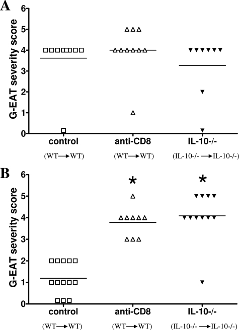
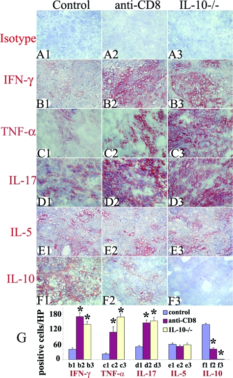
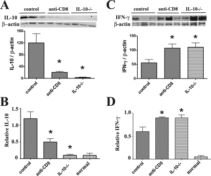
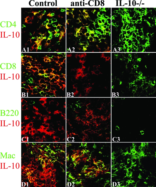
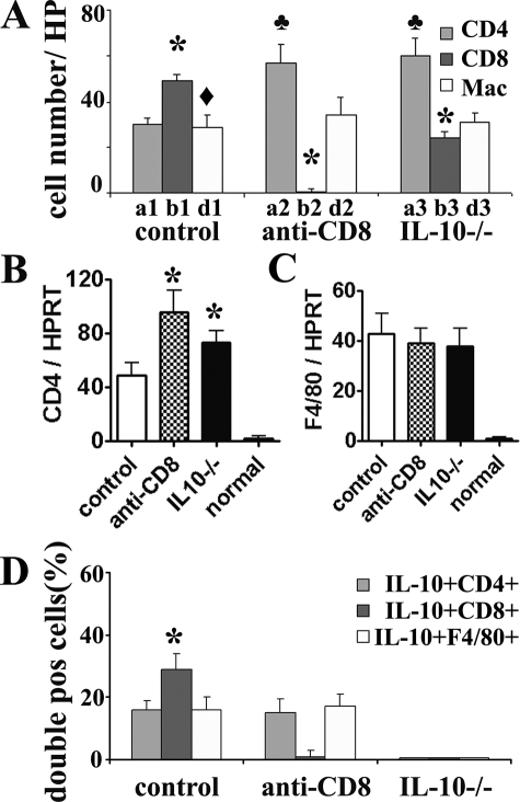
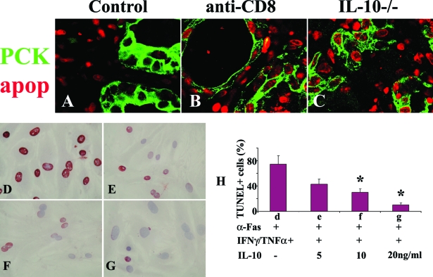
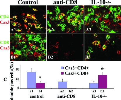
Granulomatous experimental autoimmune thyroiditis (G-EAT) is induced by mouse thyroglobulin-sensitized splenocytes activated in vitro with mouse thyroglobulin and interleukin (IL)-12. Thyroid lesions reach maximal severity 20 days after cell transfer, and inflammation either resolves or progresses to fibrosis by day 60 depending on the extent of thyroid damage at day 20. Depletion of CD8(+) T cells inhibits G-EAT resolution. Our previous studies indicated that IL-10 was generally higher in G-EAT thyroids that resolved. Using both wild-type and IL-10(-/-) CBA/J mice, this study was undertaken to determine whether G-EAT resolution would be inhibited in the absence of IL-10. The results showed that either depletion of CD8(+) T cells or IL-10 deficiency increased fibrosis and inhibited resolution of inflammation. We also found a correlation between higher expression levels of proinflammatory cytokines and preferential expression levels of proapoptotic molecules, such as FasL and TRAIL, and antiapoptotic molecules, such as FLIP and Bcl-xL, in inflammatory cells from thyroids of both CD8-depleted and IL-10-deficient mice. Furthermore, many of the CD8(+) T cells were also IL-10(+). These results suggest that IL-10 plays an important role in G-EAT resolution and might promote resolution, at least in part, through its production in CD8(+) T cells. Further understanding of the mechanisms that promote the resolution of inflammation will facilitate the development of novel strategies for treating autoimmune diseases.


Figures

Figure 1
Figure 1
G-EAT resolution is inhibited in CD8-depleted WT and IL-10−/− recipients. G-EAT severity scores of individual mice 20 and 60 days after cell transfer are shown. Control: WT (donor) to WT (recipient); anti-CD8: WT (donor) to WT (recipient) given anti-CD8; IL-10−/−: IL-10−/− (donor) to IL-10−/− (recipient). A: Thyroid lesions in all groups reached maximal severity 20 days after cell transfer, and disease severity scores were similar with an average score of 3.3 to 4.0. B: By day 60, thyroid lesions in CD8-depleted WT and IL-10−/− recipients had ongoing inflammation (average severity score, 3.8 to 4.1), whereas thyroid lesions in WT controls had begun to resolve (average severity score, 1.2). Results are pooled from two separate experiments and are representative of five experiments. A significant difference between CD8-depleted WT and IL-10−/− recipients versus WT controls is indicated by the asterisk (P < 0.001).
Figure 2
Figure 2
Effect of CD8 depletion and IL-10 deficiency on protein expression of pro- and anti-inflammatory cytokines. Isotype control staining (A1A3) was always negative. Representative thyroids in each group at day 20 expressed the proinflammatory cytokines IFN-γ (B1–B3), TNF-α (C1–C3), and IL-17 (D1–D3) and anti-inflammatory cytokines IL-5 (E1–E3) and IL-10 (F1–F3). The staining intensity for proinflammatory cytokines was stronger in thyroids of CD8-depleted WT and IL-10−/− recipients and staining intensity for IL-10 was weaker or undetectable compared to WT controls. The staining intensity for IL-5 was similar for all groups. Cytokine-positive cells (red) in five to six randomly selected high-power fields of three representative thyroids in each group were manually counted using an enlarged image. The results are summarized in G (bars b1–f3 correspond to B1–F3). A significant difference between CD8-depleted WT and IL-10−/− recipients versus WT controls is indicated by the asterisk (P < 0.05). Results are representative areas on slides from at least three individual mice examined per group with comparable G-EAT severity scores (4 to 5+). Original magnifications, ×400.
Figure 3
Figure 3
Quantitative determination of the effect of CD8 depletion and IL-10 deficiency on IL-10 and IFN-γ protein and mRNA expression At day 20, IL-10 protein was undetectable in thyroids of IL-10−/− recipients of IL-10−/− splenocytes (A) and IL-10 was lower in thyroids of CD8-depleted WT recipients compared to WT controls. Real-time quantitative PCR indicated that IL-10 mRNA was undetectable in thyroids of IL-10−/− recipients of IL-10−/− splenocytes (B) and IL-10 mRNA was lower in thyroids of CD8-depleted recipients compared to WT controls. IFN-γ protein (C) and mRNA (D) expression was higher in thyroids of CD8-depleted WT or IL-10−/− recipients compared to WT controls. For Western blot, 30 μg of protein from thyroids of each group (three individuals) was loaded in each lane and results are shown at the top in A and C. Results are expressed as the mean ratio of densitometric U/β-actin ± SEM (×100), and are representative of two independent experiments. For real-time PCR, bars are means of data for thyroids of four to five individual mice ± SEM. Results are expressed as the mean relative ratio to HPRT. A significant difference between CD8-depleted WT and IL-10−/− recipients versus WT controls is indicated by the asterisk (P < 0.05).
Figure 4
Figure 4
Cell distribution of IL-10 protein in thyroids of CD8-depleted recipients and WT controls. Dual-color immunofluorescence and confocal laser-scanning microscopy on thyroid frozen sections 20 days after cell transfer. CD4+ and CD8+ T cells, B cells, and macrophages were identified by cell surface expression of CD4, CD8, B220, or F4/80 (A1–D3, green), and IL-10-producing cells were identified by IL-10 staining (red). IL-10 was undetectable in thyroids of IL-10−/− recipients (A3–D3, red) and few CD8+ T cells were detected in CD8-depleted WT recipients (B2, green). IL-10 was produced by CD4+ T cells (A1, yellow, overlay), CD8+ T cells (B1, yellow, overlay), and macrophages (D1, yellow, overlay) in thyroids of WT controls. After depletion of CD8+ T cells, most IL-10 was produced by CD4+ T cells (A2, yellow, overlay) and macrophages (D2, yellow, overlay). No IL-10+ B cells were detected (C1–C3). CD8+ T cells (B1, yellow plus green) outnumbered CD4+ T cells (A1, yellow plus green) in WT controls, whereas CD4+ T cells outnumbered CD8+ T cells in CD8-depleted WT or IL-10−/− recipients (A1–A3 versus B1–B3). Shown are representative areas on slides of thyroids of at least three individual mice per group with comparable 4 to 5+ G-EAT severity scores. Original magnifications, ×800.
Figure 5
Figure 5
Effect of CD8 depletion on the percentages of IL-10-producing CD4+ T cells and macrophages. Numbers of CD4+, CD8+ T cells, and macrophages in five to six randomly selected high-power fields shown in Figure 4 (original magnifications, ×800) were manually counted and summarized in A (bars a1–d3 correspond to A1–D3 in Figure 4). A significant difference between CD8+ and CD4+ T cells in each group is indicated by the asterisk (P < 0.05). A significant difference between CD8+ T cells and macrophages in WT controls is indicated by the diamond (P < 0.05). A significant difference of CD4+ T cells in each group is indicated by the club (P < 0.05). mRNA was isolated from individual thyroid lobes 20 days after cell transfer and amplified as described in the Materials and Methods. B and C: Expression of CD4 and F4/80 mRNA is shown for each group including normal (naïve) mice. Results are expressed as the mean ratio of CD4 or F4/80 densitometric U/HPRT ± SEM (×100) of five to six mice per group, and are representative of two independent experiments. A significant difference between CD8-depleted WT or IL-10−/− recipients and WT controls is indicated by the asterisk (P < 0.05). D: IL-10+CD4+ cells (yellow, overlay in Figure 4, A1–A3), IL-10+CD8+ cells (yellow, overlay in Figure 4, B1–B3), and IL-10+F4/80+ cells (yellow, overlay in Figure 4, D1–D3) were manually counted in three to four randomly selected low-power fields (original magnifications, ×200) and expressed as the percentage of total CD4+ cells, CD8+ cells, or F4/80+ cells and summarized. The percentage of IL-10+CD8+ cells in thyroids of WT controls is significantly higher than that of IL-10+CD4+ or IL-10+F4/80+ cells in WT controls (P < 0.05).
Figure 6
Figure 6
IL-10 protects TECs from Fas-mediated apoptosis in vitro and in vivo. A–C: The distribution pattern of apoptotic cells in each group was determined by a fluorescence apoptosis kit. TECs were identified by pan-cytokeratin (PCK, green) and apoptosis was detected as red nuclear staining. Most apoptotic cells (red) were inflammatory cells in WT controls (A) and most were TECs in thyroids of CD8-depleted WT (B) and IL-10−/− recipients (C). Shown are representative areas on slides of thyroids of at least three individual mice per group with comparable G-EAT severity scores. Sixty to seventy percent confluent cultured TECs were pretreated with IFN-γ/TNF-α for 4 days in the presence (5 to 20 ng/ml) or absence of IL-10 and stimulated with anti-Fas for 20 hours. Apoptosis was detected by TUNEL staining. TUNEL+ cells (D–G, red) in five to six randomly selected high-power fields of three individual mice per group were manually counted and summarized in H (bars d–g correspond to D–G). In the absence of IL-10, TECs cultured with cytokines and anti-Fas underwent extensive Fas-mediated apoptosis (74 ± 13% TUNEL+ cells), whereas the percentage of TUNEL+ cells decreased in the presence of IL-10 in a dose-dependent manner. A significant difference in the percentage of TUNEL+ cells in the presence of different concentrations of IL-10 compared to no IL-10 is indicated by the asterisk (P < 0.05). Original magnifications: ×800 (A–C); ×400 (D–G).
Figure 7
Figure 7
CD8+ T cells in thyroids of mice with G-EAT are protected from apoptosis by IL-10. Confocal staining of CD4 and CD8 on frozen sections with active caspase-3 at day 20. Active caspase-3 (red, A1–B3) is positive in both CD4+ (green, A1–A3) and CD8+ (green, B1–B3) T cells (yellow, overlay). Apoptotic (active caspase-3+) inflammatory cells in controls are mainly CD4+ T cells (A1, yellow, overlay), whereas most apoptotic cells in IL-10−/− recipients of IL-10−/− donor cells are CD8+ T cells (B3, yellow, overlay). Active caspase-3+CD4+ or active caspase-3+CD8+ cells were manually counted in five to six randomly selected high-power fields and expressed as the percentage of total CD4+ or CD8+ T cells and summarized in C. Shown are representative areas on slides of thyroids of at least three individual mice per group with comparable 4 to 5+ G-EAT severity scores. A significant difference between the percentage of active caspase-3+CD4+ and active caspase-3+CD8+ cells in WT controls or in IL-10−/− group is indicated by the asterisk (P < 0.05). Original magnifications, ×800.

Similar articles

Cited by

References

    1. Conaway DH, Giraldo AA, David CS, Kong YC. In situ analysis of T cell subset composition in experimental autoimmune thyroiditis after adoptive transfer of activated spleen cells. Cell Immunol. 1990;125:247–253. - PubMed
    1. Lira SA, Martin AP, Marinkovic T, Furtado GC. Mechanisms regulating lymphocytic infiltration of the thyroid in murine models of thyroiditis. Crit Rev Immunol. 2005;25:251–262. - PubMed
    1. Braley-Mullen H, Johnson M, Sharp GC, Kyriakos M. Induction of experimental autoimmune thyroiditis in mice with in vitro activated splenic T cells. Cell Immunol. 1985;93:132–143. - PubMed
    1. Braley-Mullen H, Sharp GC, Bickel JT, Kyriakos M. Induction of severe granulomatous experimental autoimmune thyroiditis in mice by effector cells activated in the presence of anti-interleukin 2 receptor antibody. J Exp Med. 1991;173:899–912. - PMC - PubMed
    1. Braley-Mullen H, Sharp GC, Tang H, Chen K, Kyriakos M, Bickel JT. Interleukin-12 promotes activation of effector cells that induce a severe destructive granulomatous form of murine experimental autoimmune thyroiditis. Am J Pathol. 1998;152:1347–1358. - PMC - PubMed

Publication types

MeSH terms