. 2008 Aug;104(2):261-73.
doi: 10.1093/toxsci/kfn093. Epub 2008 May 8.

Acquired resistance to acetaminophen hepatotoxicity is associated with induction of multidrug resistance-associated protein 4 (Mrp4) in proliferating hepatocytes

Affiliations

Acquired resistance to acetaminophen hepatotoxicity is associated with induction of multidrug resistance-associated protein 4 (Mrp4) in proliferating hepatocytes

Lauren M Aleksunes et al. Toxicol Sci. 2008 Aug.

Abstract

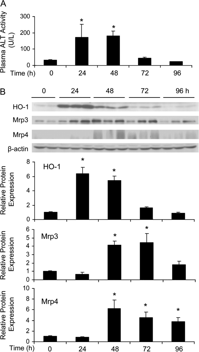
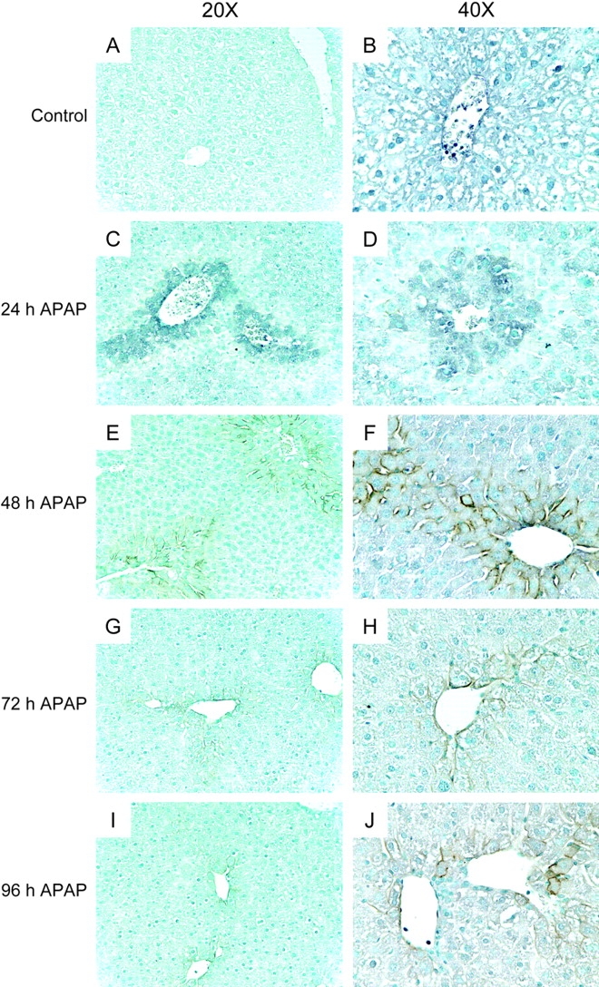
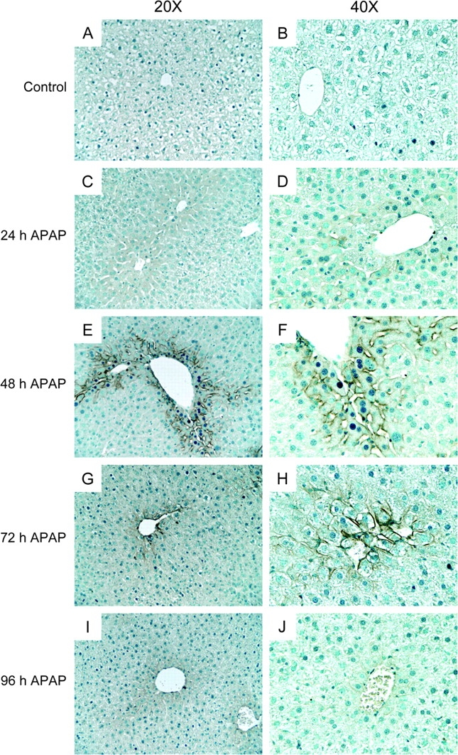
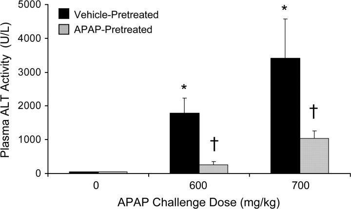
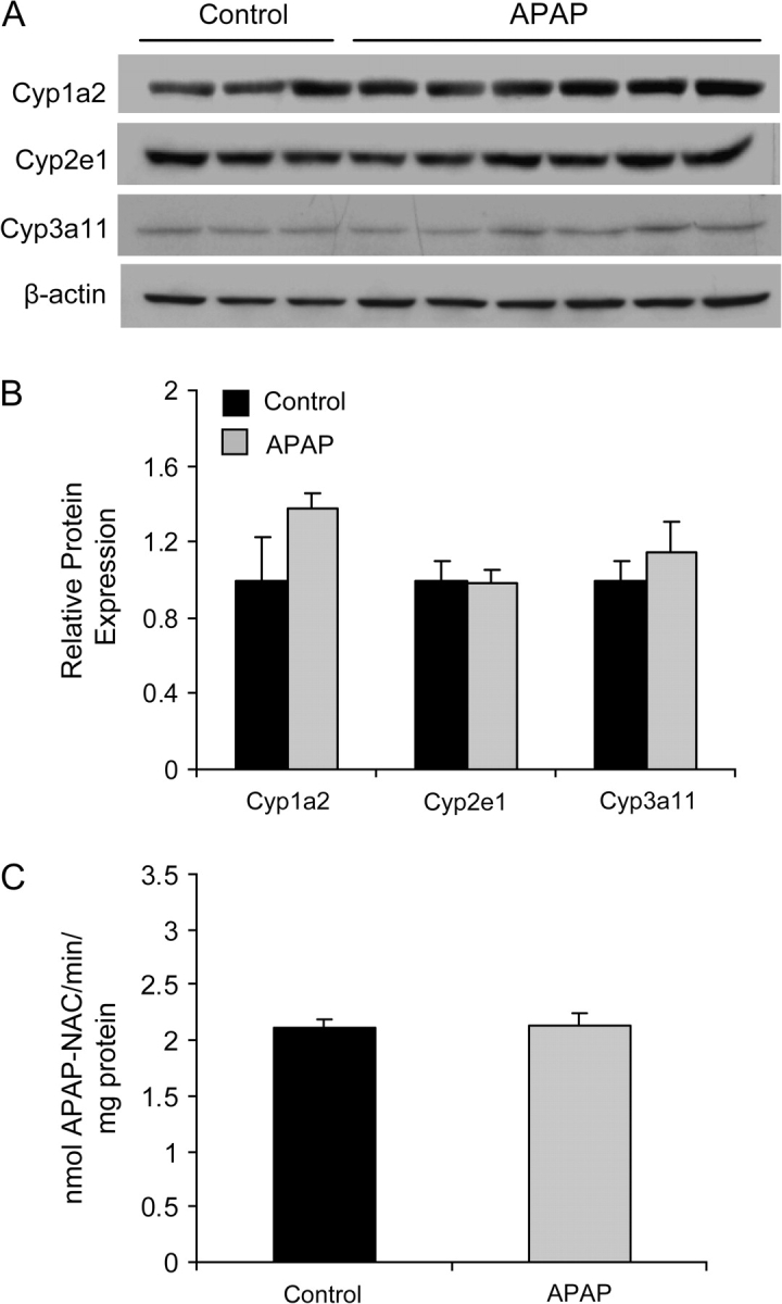
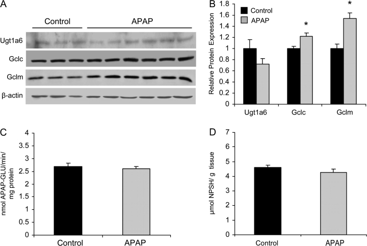
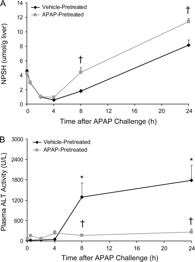
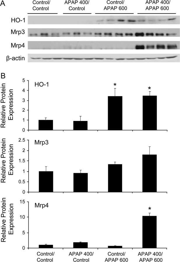
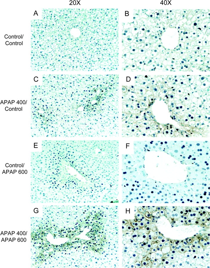
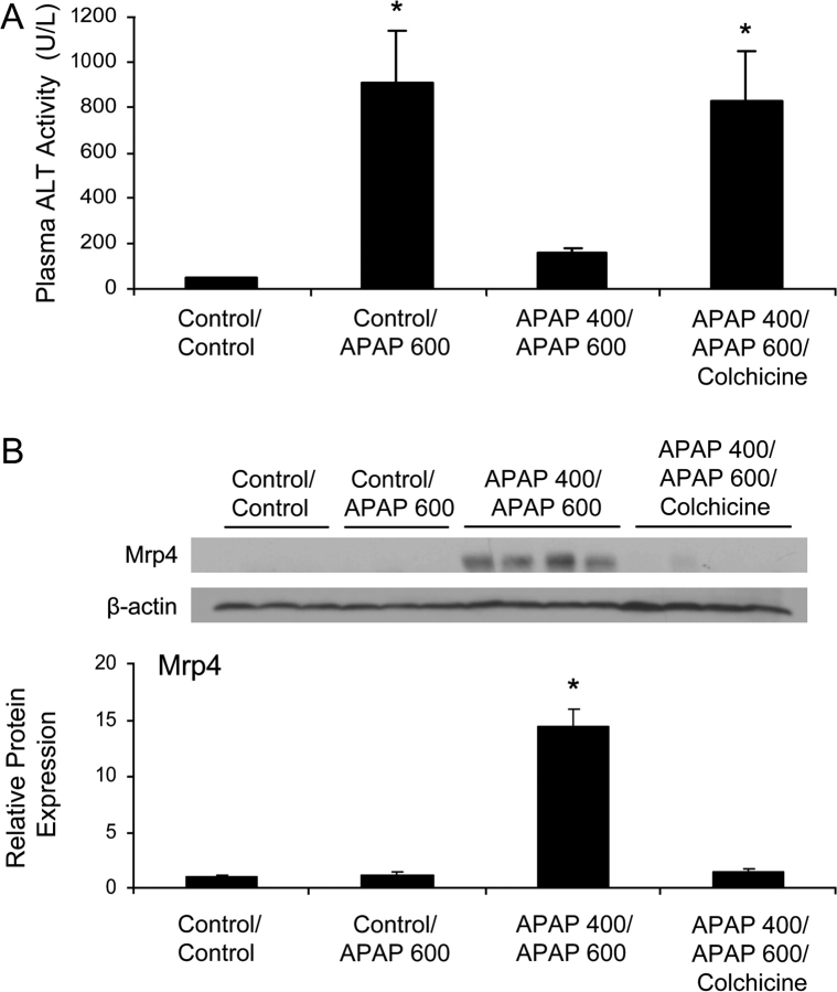
Treatment with hepatotoxicants such as acetaminophen (APAP) causes resistance to a second, higher dose of the same toxicant (autoprotection). APAP induces hepatic mRNA and protein levels of the multidrug resistance-associated proteins (Mrp) transporters in mice and humans. Basolateral efflux transporters Mrp3 and Mrp4 are the most significantly induced. We hypothesized that upregulation of Mrp3 and Mrp4 is one mechanism by which hepatocytes become resistant to a subsequent higher dose of APAP by limiting accumulation of xeno-, endobiotics, and byproducts of hepatocellular injury. The purpose of this study was to evaluate Mrp3 and Mrp4 expression in proliferating hepatocytes in a mouse model of APAP autoprotection. Plasma and livers were collected from male C57BL/6J mice treated with APAP 400 mg/kg for determination of hepatotoxicity and protein expression. Maximal Mrp3 and Mrp4 induction occurred 48 h after APAP. Mrp4 upregulation occurred selectively in proliferating hepatocytes. Additional groups of APAP-pretreated mice were challenged 48 h later with a second, higher dose of APAP. APAP-pretreated mice had reduced hepatotoxicity after APAP challenge compared to those pretreated with vehicle. A more rapid recovery of glutathione (GSH) in APAP-pretreated mice corresponded with increases in GSH synthetic enzymes. Interestingly, mice pretreated and challenged with APAP had dramatic increases in Mrp4 expression as well as enhanced hepatocyte proliferation. Inhibition of hepatocyte replication with colchicine not only restored sensitivity of APAP-pretreated mice to injury, but also blocked Mrp4 induction. Mrp4 overexpression may be one phenotypic property of proliferating hepatocytes that protects against subsequent hepatotoxicant exposure by mechanisms that are presently unknown.


Figures

FIG. 1.
FIG. 1.
Plasma ALT activity and HO-1, Mrp3, Mrp4 protein expression after APAP treatment. Plasma and livers were isolated from mice 24, 48, 72, and 96 h following APAP 400 mg/kg or vehicle administration. (A) The data are presented as mean plasma ALT activity (U/l) ± SE (n = 4–8 animals). (B) Western blots for HO-1, Mrp3, and Mrp4 were performed using liver membrane preparations from control and APAP-treated mice. Equal protein loading (50 μg protein/lane) was confirmed by detection of β-actin. Representative time course blots (n = 2–3 animals) are shown (upper panel). The data from individual Western blots (for each time point) are presented as mean relative protein expression ± SE (n = 4–6 animals) (lower panel). Asterisks (*) represent a statistical difference (p < 0.05) between pooled 0-h vehicle- and APAP-treated groups.
FIG. 2.
FIG. 2.
Immunohistochemical detection of 4-HNE in relation to Mrp4 after APAP treatment. Immunohistochemical staining to detect 4-HNE (purple) and Mrp4 (brown) was conducted on paraffin-embedded liver sections from control and APAP 400 mg/kg-treated mice at 24, 48, 72, and 96 h. Representative images are shown at two magnifications (20× and 40×). (A/B) Control; (C/D) APAP-treated 24 h; (E/F) APAP-treated 48 h; (G/H) APAP-treated 72 h; (I/J) APAP-treated 96-h liver sections.
FIG. 3.
FIG. 3.
Immunohistochemical detection of PCNA in relation to Mrp4 after APAP treatment. Immunohistochemical staining to detect PCNA (purple) and Mrp4 (brown) was conducted on paraffin-embedded liver sections from control and APAP 400 mg/kg-treated mice at 24, 48, 72, and 96 h. Representative images are shown at two magnifications (20× and 40×). Panels (A/B) control; (C/D) APAP-treated 24 h; (E/F) APAP-treated 48 h; (G/H) APAP-treated 72 h; (I/J) APAP-treated 96-h liver sections.
FIG. 4.
FIG. 4.
Plasma ALT activity after APAP challenge in vehicle- and APAP-pretreated mice. Groups of mice were treated with vehicle or APAP 400 mg/kg. Forty-eight h later mice were challenged with vehicle or APAP (600 or 700 mg/kg). Plasma was isolated from mice 24 h following APAP challenge. The data are presented as mean plasma ALT activity (U/l) ± SE (n = 4–8 animals). Asterisks (*) represent a statistical difference (p < 0.05) between vehicle- and APAP-treated groups. Daggers (†) represent a statistical difference between vehicle-pretreated, APAP-challenged and APAP-pretreated, APAP-challenged groups.
FIG. 5.
FIG. 5.
Bioactivation pathways for APAP in livers from vehicle- and APAP-pretreated mice. Western blots for Cyp1a2, Cyp2e1 and Cyp3a11 proteins were performed using homogenate preparations 48 h after vehicle (Control) or APAP 400 mg/kg treatment. The data are presented as individual blots (A) and as mean relative protein expression ± SE (n = 3–6 animals) (B). Equal protein loading (50 μg protein/lane) was confirmed by detection of β-actin. (C) Microsomes were prepared and assayed for APAP bioactivation as described in “Materials and Methods” (n = 4 animals). Data are presented as nmol APAP-NAC/min/mg protein. Asterisks (*) represent a statistical difference (p < 0.05) between control and APAP-treated groups.
FIG. 6.
FIG. 6.
Detoxification pathways for APAP in livers from vehicle- and APAP-pretreated mice. Western blots for Ugt1a6, Gclc, and Gclm proteins were performed using homogenate preparations 48 h after vehicle (Control) or APAP 400 mg/kg treatment. The data are presented as individual blots (A) and as mean relative protein expression ± SE (B). Equal protein loading (50 μg protein/lane) was confirmed by detection of β-actin. (C) Microsomes were prepared and assayed for APAP glucuronidation as described in Materials and Methods. Data are presented as nmol APAP-GLU/min/mg protein. (D) Basal NPSH levels expressed as μmol NPSH/g tissue. Asterisks (*) represent a statistical difference (p < 0.05) between control and APAP-treated groups.
FIG. 7.
FIG. 7.
GSH recovery in livers after APAP pretreatment and challenge. Plasma and liver were collected from vehicle- and APAP-pretreated mice at 0.5, 2, 4, 8, and 24 h after APAP 600 mg/kg challenge. (A) The data are presented as mean NPSH (μmol/g liver) ± SE (n = 4 animals). (B) The data are presented as mean plasma ALT activity (U/l) ± SE (n = 4 animals). Asterisks (*) represent a statistical difference (p < 0.05) between vehicle- and APAP-treated groups. Daggers (†) represent a statistical difference between vehicle-pretreated, APAP-challenged and APAP-pretreated, APAP-challenged groups.
FIG. 8.
FIG. 8.
HO-1, Mrp3, and Mrp4 protein expression after APAP pretreatment and challenge. Groups of mice were treated with vehicle (Control) or APAP (400 mg/kg: APAP 400). Forty-eight hours later mice were challenged with vehicle (Control) or APAP (600 mg/kg: APAP 600). Livers were isolated from mice 24 h following APAP challenge. Western blots for HO-1, Mrp3, and Mrp4 were performed using liver membrane preparations. Equal protein loading (50 μg protein/lane) was confirmed by detection of β-actin. (A) The data are presented as representative blots (n = 3) and (B) as mean relative protein expression ± SE (n = 3). Asterisks (*) represent a statistical difference (p < 0.05) from Control/Control mice.
FIG. 9.
FIG. 9.
Immunohistochemical detection of Mrp4 and PCNA after APAP pretreatment and challenge. Groups of mice were treated with vehicle (Control) or APAP (400 mg/kg: APAP 400). Forty-eight hours later mice were challenged with vehicle (Control) or APAP (600 mg/kg: APAP 600). Livers were isolated from mice 24 h following APAP challenge. Immunohistochemical staining to detect PCNA (purple) and Mrp4 (brown) was conducted on paraffin-embedded liver sections. Representative images are shown at two magnifications (20× and 40×). Panels (A/B) Control/Control; (C/D) APAP 400/Control; (E/F) Control/APAP 600; (G/H) APAP 400/APAP 600 liver sections.
FIG. 10.
FIG. 10.
Plasma ALT activity and Mrp4 protein expression after colchicine intervention in APAP autoprotected mice. Groups of mice were treated with vehicle (Control) or APAP (400 mg/kg: APAP 400). Colchicine (2 mg/kg) was administered at 24 and 49 h after APAP 400 mg/kg. Mice were challenged with vehicle (Control) or APAP (600 mg/kg: APAP 600) at 48 h. Plasma and livers were isolated from mice 24 h following APAP challenge. (A) The data are presented as mean plasma ALT activity (U/l) ± SE (n = 4–8 animals). (B) Western blot for Mrp4 was performed using liver membrane preparations. Equal protein loading (50 μg protein/lane) was confirmed by detection of β-actin. Asterisks (*) represent a statistical difference (p < 0.05) from Control/Control mice.

Similar articles

Cited by

References

    1. Aleksunes LM, Scheffer GL, Jakowski AB, Pruimboom-Brees IM, Manautou JE. Coordinated expression of multidrug resistance-associated proteins (Mrps) in mouse liver during toxicant-induced injury. Toxicol. Sci. 2006;89:370–379. - PubMed
    1. Aleksunes LM, Slitt AM, Cherrington NJ, Thibodeau MS, Klaassen CD, Manautou JE. Differential expression of mouse hepatic transporter genes in response to acetaminophen and carbon tetrachloride. Toxicol. Sci. 2005;83:44–52. - PubMed
    1. Aleksunes LM, Slitt AL, Maher JM, Augustine LM, Goedken MJ, Chan JY, Cherrington NJ, Klaassen CD, Manautou JE. Induction of Mrp3 and Mrp4 transporters during acetaminophen hepatotoxicity is dependent on Nrf2. Toxicol. Appl. Pharmacol. 2008;226:74–83. - PMC - PubMed
    1. Bai J, Lai L, Yeo HC, Goh BC, Tan TM. Multidrug resistance protein 4 (MRP4/ABCC4) mediates efflux of bimane-glutathione. Int. J. Biochem. Cell. Biol. 2004;36:247–257. - PubMed
    1. Bajt ML, Knight TR, Farhood A, Jaeschke H. Scavenging peroxynitrite with glutathione promotes regeneration and enhances survival during acetaminophen-induced liver injury in mice. J. Pharmacol. Exp. Ther. 2003;307:67–73. - PubMed

Publication types

MeSH terms