. 2008 Jul 25;283(30):20754-60.
doi: 10.1074/jbc.M800024200. Epub 2008 May 9.

Phosphorylation of Bad at Thr-201 by JNK1 promotes glycolysis through activation of phosphofructokinase-1

Affiliations

Phosphorylation of Bad at Thr-201 by JNK1 promotes glycolysis through activation of phosphofructokinase-1

Hongbin Deng et al. J Biol Chem. .

Abstract

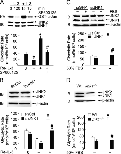
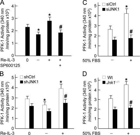
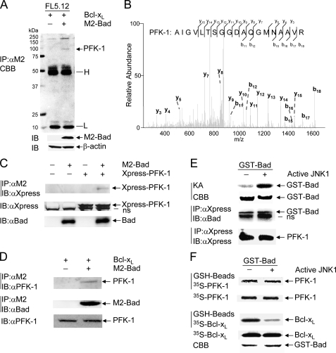
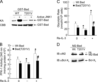
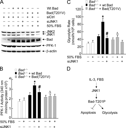
The mitogen-activated protein kinase JNK1 suppresses interleukin-3 withdrawal-induced cell death through phosphorylation of the BH3-only pro-apoptotic Bcl-2 family protein Bad at Thr-201. It is unknown whether JNK1 regulates glycolysis, an important metabolic process that is involved in cell survival, and if so, whether the regulation depends on Thr-201 phosphorylation of Bad. Here we report that phosphorylation of Bad by JNK1 is required for glycolysis through activation of phosphofructokinase-1 (PFK-1), one of the key enzymes that catalyze glycolysis. Genetic disruption of Jnk1 alleles or silencing of Jnk1 by small interfering RNA abrogates glycolysis induced by growth/survival factors such as serum or interleukin-3. Proteomic analysis identifies PFK-1 as a novel Bad-associated protein. Although the interaction between PFK-1 and Bad is independent of JNK1, Thr-201 phosphorylation of Bad by JNK1 is required for PFK-1 activation. Thus, our results provide a novel molecular mechanism by which JNK1 promotes glycolysis for cell survival.


Figures

FIGURE 1.
FIGURE 1.
JNK1 is essential for IL-3- or serum-induced glycolysis. A, FL5.12 Bcl-XL/Bad (FL-5.12) cells were deprived of IL-3 for 2 h (–IL-3) followed by IL-3 readdition (+IL-3) for 15 min. The specific JNK inhibitor SP600125 (10 μm) was added 30 min prior to IL-3 readdition (Re-IL-3). The glycolytic rate (*, p < 0.01 when compared with 0 h; #, p < 0.01 when compared with IL-3 readdition for 15 min) was determined by measuring the conversion of [5-3H]glucose to [3H]H2O, as described under “Materials and Methods.” The results are presented as means ± S.E. and represent three individual experiments. The JNK activity was measured by immune complex kinase assays (KA) using GST-c-Jun as substrate, and the expression level of JNK was detected by immunoblotting (IB) using anti-JNK antibody (Pharmingen, 666.6). IP, immunoprecipitation. B, FL-5.12 cells were infected by retroviral vectors encoding the control shRNA (shCtrl) or shRNA of JNK1 (shJNK1). Cells were deprived of IL-3 for 2 h (–IL-3) followed by IL-3 readdition (+IL-3) for 15 min. The effect of shRNA of JNK1 on JNK1 expression was detected by immunoblotting and the glycolytic rate (*, p < 0.01 when compared with 0 h; #, p < 0.01 when compared with control shRNA cells treated with IL-3 readdition for 15 min) was determined, as described in panel A. C, immortalized WT MEFs were transfected with siCtrl or siJNK1 (200 nm each) followed by stimulation without or with 50% FBS for 15 min. The glycolytic rate (*, p < 0.01 when compared with unstimulated siCtrl cells; #, p < 0.01 when compared with siCtrl cells treated with 50% FBS stimulation for 15 min) was determined as described in panel A. D, WT or Jnk1–/– MEFs were stimulated without or with 50% FBS for 15 min, and the glycolytic rate (*, p < 0.01 when compared with unstimulated WT MEFs; #, p < 0.01 when compared with WT MEFs treated with 50% FBS stimulation for 15 min) was determined as described in panel A. ns, nonspecific.
FIGURE 2.
FIGURE 2.
JNK1 regulates the activation of PFK-1. A, FL-5.12 cells were deprived of IL-3 for 2 h (–IL-3) followed by IL-3 readdition (Re-IL-3, +IL-3) for 15 min. The specific JNK inhibitor SP600125 (10 μm) was added 30 min prior to IL-3 readdition. Cell lysates were analyzed for PFK-1 activity (*, p < 0.05 when compared with 0 h; #, p < 0.01 when compared with IL-3 readdition for 15 min), as described under “Materials and Methods.” B, FL-5.12 cells were infected by control shRNA (shCtrl) or shRNA of JNK1 (shJNK1) retrovirus. Cells were deprived of IL-3 for 2 h (–IL-3) followed by IL-3 readdition (+IL-3) for 15 min. PFK-1 activity (*, p < 0.05 when compared with 0 h; #, p < 0.05 when compared with shCtrl cells treated with IL-3 readdition for 15 min) was measured as described in panel A. C, WT MEFs were transfected with siCtrl or siJNK1 (200 nm each) followed by stimulation without or with 50% FBS for 15 min. PFK-1 activity (*, p < 0.05 when compared with unstimulated siCtrl cells; #, p < 0.01 when compared with siCtrl cells treated with 50%FBS stimulation for 15 min) was determined as described in panel A. D, WT or Jnk1–/– MEFs were stimulated without or with 50% FBS for 15 min. PFK-1 activity (*, p < 0.01 when compared with unstimulated WT MEFs; #, p < 0.01 when compared with WT MEFs treated with 50%FBS stimulation for 15 min) was determined as described in panel A.
FIGURE 3.
FIGURE 3.
PFK-1 is a novel Bad-associated protein. A, cell extracts of FL5.12/Bcl-XL and FL-5.12 were subjected to immunoprecipitation with anti-M2 antibody, followed by SDS-PAGE, and stained with colloidal Coomassie Brilliant Blue (CBB). Transfected M2-Bad was analyzed by immunoblotting using anti-M2 antibody. The arrow indicates the major Bad-associated protein, which was identified as PFK-1 (see panel B below). H, IgG heavy chains; L, IgG light chains. B, the major Bad-associated protein identified in panel A was excised and sequenced by nano-HPLC electrospray ion trap mass spectrometry, as described under “Materials and Methods.” C, COS-1 cells were transfected with mammalian expression vectors encoding PFK-1 or M2-Bad (2 μgof each), without or with empty vector (2 μg). After 40 h, M2-Bad was immunoprecipitated (IP) with anti-M2 antibody, and M2-Bad-associated PFK-1 was immunoblotted (IB) with anti-Xpress antibody. Total M2-Bad or Xpress-PFK-1 proteins in the cell extracts were analyzed by immunoblotting with anti-Bad or anti-Xpress antibody, respectively. D, M2-Bad was immunoprecipitated from stable FL5.12/Bcl-XL or FL-5.12 cells with anti-M2 antibody, and M2-Bad-associated PFK-1 was analyzed by immunoblotting using anti-PFK-1 antibody. The amounts of immunoprecipitated M2-Bad proteins and total PFK-1 proteins were examined by immunoblotting with anti-Bad and anti-PFK-1 antibody, respectively. E, purified GST-Bad proteins were incubated without or with active JNK1 in a kinase reaction buffer in the presence of [γ-32P] ATP (top two panels, KA for kinase assays) or 17 μm nonradioactive ATP (low two panels). GST-Bad proteins modified with nonradioactive ATP were mixed with cell extracts of COS-1 cells that have been transfected with mammalian expression vector encoding Xpress-PFK-1. Xpress-PFK-1 was immunoprecipitated with anti-Xpress antibody, and Xpress-PFK-1-associated GST-Bad proteins were analyzed by immunoblotting using anti-Bad antibody. The amount of Xpress-PFK-1 in the immune complex was analyzed by immunoblotting with anti-Xpress antibody. F, GST-Bad proteins modified with nonradioactive ATP (as described in panel E) were mixed with in vitro translated 35S-labeled PFK-1 or 35S-labeled Bcl-XL for 12 h and extensively washed. GST-Bad-associated 35S-PFK-1 or 35S-Bcl-XL was visualized by radioautography. One-tenth of the input 35S-PFK-1 or 35S-Bcl-XL was analyzed by radioautography. The amount of GST-Bad proteins was examined by Coomassie Brilliant Blue staining. ns, nonspecific.
FIGURE 4.
FIGURE 4.
JNK1-phosphorylated Bad regulates PFK-1 activation and glycolysis. A, purified WT GST-Bad and GST-Bad(T201V) mutant proteins were incubated with active JNK1 for 60 min, and phosphorylation of GST-Bad proteins was measured by in vitro kinase assays (KA) in the presence of [γ-32P]ATP. The amount of GST-Bad was examined by Coomassie Brilliant Blue (CBB) staining. B and C, FL5.12/Bcl-XL cells were transfected with WT M2-Bad or M2-Bad(T201V) (20 μg of each). 48 h later, cells were deprived of IL-3 for 2 h (–IL-3) followed by IL-3 readdition (Re-IL-3, +IL-3) for 15 min. PFK-1 activity (B, *, p < 0.05 when compared with cells transfected with WT M2-Bad at 0 h; #, p < 0.05 when compared with cells transfected with WT M2-Bad treated with IL-3 readdition for 15 min) and the glycolytic rate (C, *, p < 0.01 when compared with cells transfected with WT M2-Bad at 0 h; #, p < 0.01 when compared with cells transfected with WT M2-Bad treated with IL-3 readdition for 15 min) were measured as described in the legends for Figs. 1 and 2. D, the expression levels of Bad and Bcl-XL in transfected FL5.12/Bcl-XL cells were analyzed by immunoblotting using anti-M2 and anti-Bcl-XL antibody, respectively. IB, immunoblotting; IP, immunoprecipitation.
FIGURE 5.
FIGURE 5.
JNK1-mediated activation of PFK-1 and glycolysis depends on its phosphorylation of Bad at Thr-201. A, Bad–/– cells were transfected with mammalian expressing vector encoding WT M2-Bad, M2-Bad(T201V), or empty vector (2μg of each), without or with siCtrl or siJNK1 (200 nm each). The expression levels of JNK, Bad, and PFK-1 were analyzed by immunoblotting with the antibodies against JNK, Bad, or PFK-1, respectively. B and C, after 48 h, cells were treated without or with 50% FBS for 15 min. The activity of PFK-1 (B, *, p < 0.01 when compared with untreated cells transfected with WT M2-Bad; #, p < 0.01 and ▵, p < 0.05 when compared with cells transfected with WT M2-Bad treated with 50% FBS stimulation for 15 min, respectively) and the glycolytic rate (C, *, p < 0.01 when compared with untreated cells transfected with WT M2-Bad; # and ▵, p < 0.01 when compared with cells transfected with WT M2-Bad treated with 50% FBS stimulation for 15 min) were measured as described in the legends for Figs. 1 and 2. D, the schematic presentation of how JNK1 regulates PFK-1 activity via phosphorylation of Bad at Thr-201, thereby contributing to glycolysis.

References

    1. Hibi, M., Lin, A., Smeal, T., Minden, A., and Karin, M. (1993) Genes Dev. 7 2135–2148 - PubMed
    1. Chang, L., and Karin, M. (2001) Nature 410 37–40 - PubMed
    1. Davis, R. J. (2000) Cell 103 239–252 - PubMed
    1. Lin, A. (2003) BioEssays 25 17–24 - PubMed
    1. Liu, J., Minemoto, Y., and Lin, A. (2004) Mol. Cell Biol. 24 10844–10856 - PMC - PubMed

Publication types

LinkOut - more resources