Review
. 2008 Jul;93(7):773-97.
doi: 10.1113/expphysiol.2007.041848. Epub 2008 May 9.

Central control of thermogenesis in mammals

Affiliations
Review

Central control of thermogenesis in mammals

Shaun F Morrison et al. Exp Physiol. 2008 Jul.

Abstract

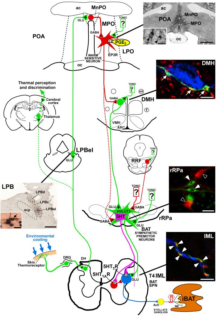
Thermogenesis, the production of heat energy, is an essential component of the homeostatic repertoire to maintain body temperature in mammals and birds during the challenge of low environmental temperature and plays a key role in elevating body temperature during the febrile response to infection. The primary sources of neurally regulated metabolic heat production are mitochondrial oxidation in brown adipose tissue, increases in heart rate and shivering in skeletal muscle. Thermogenesis is regulated in each of these tissues by parallel networks in the central nervous system, which respond to feedforward afferent signals from cutaneous and core body thermoreceptors and to feedback signals from brain thermosensitive neurons to activate the appropriate sympathetic and somatic efferents. This review summarizes the research leading to a model of the feedforward reflex pathway through which environmental cold stimulates thermogenesis and discusses the influence on this thermoregulatory network of the pyrogenic mediator, prostaglandin E(2), to increase body temperature. The cold thermal afferent circuit from cutaneous thermal receptors ascends via second-order thermosensory neurons in the dorsal horn of the spinal cord to activate neurons in the lateral parabrachial nucleus, which drive GABAergic interneurons in the preoptic area to inhibit warm-sensitive, inhibitory output neurons of the preoptic area. The resulting disinhibition of thermogenesis-promoting neurons in the dorsomedial hypothalamus and possibly of sympathetic and somatic premotor neurons in the rostral ventromedial medulla, including the raphe pallidus, activates excitatory inputs to spinal sympathetic and somatic motor circuits to drive thermogenesis.


Figures

Figure 1
Figure 1. Schematic diagram summarizing the proposed model for the feedforward reflex circuit for cold-evoked brown adipose tissue thermogenesis
Photomicrographs of the lateral parabrachial nucleus (LPB), the preoptic area (POA), the dorsomedial hypothalamus (DMH), the rostral raphe pallidus (rRPa) and spinal intermediolateral nucleus (IML) illustrate the anatomical substrates for key neurochemical and synaptic aspects of the proposed thermoregulatory circuit. In the LPB panel, (LPBel), central (LPBc) and dorsal (LPBd) subnuclei of the LPB contain neurons that are retrogradely labelled with a tracer (brown) from the median preoptic (MnPO) subregion of the POA. Retrogradely labelled neurons in the LPBel and LPBc, but not those in the LPBd, also express Fos (blue-black nuclei) following cold exposure of the animals (arrow, inset); scp, superior cerebellar peduncle; scale bars represent 0.5 mm and 15 μm (inset). Reproduced with permission from Nakamura & Morrison, 2008b). In the POA panel, immunohistochemistry for EP3 receptors (EP3R) shows the localization of these receptors in cell bodies (inset, arrowheads) and dendritic fibres of neurons that are distributed in the MnPO and medial preoptic (MPO) subregions of the POA; ac, anterior commissure; oc, optic chiasm; LPO, lateral preoptic area; PGE2, prostaglandin E2; GLU, glutamate; scale bars represent 1 mm and 20 μm (inset). Reproduced with permission from Nakamura et al. (1999). In the DMH panel, axon swellings of POA neurons (green) that are positive for a marker of GABAergic terminals (red) are closely apposed (arrows) to neurons that are retrogradely labelled with a tracer (blue) from the rRPa; ARC, arcuate nucleus; f, fornix; mt, mammillothalamic tract; VMH, ventromedial hypothalamic nucleus; RRF, retrorubral field; scale bar represents 5 μm. Reproduced with permission from Nakamura et al. (2005). The rRPa panel shows double immunofluorescence labelling for vesicular glutamate transporter 3 (VGLUT3)-positive (green, white arrowheads) and serotonin (5-HT)-positive neurons (red, open arrowheads); BAT, brown adipose tissue; scale bar represents 30 μm. The IML panel shows that axon swellings of rRPa neurons (green) that are positive for VGLUT3 (red) are closely associated (white arrowheads) with dendritic fibres positive (blue) for a marker of sympathetic preganglionic neurons (SPNs); Ach, acetylcholine; DH, dorsal horn; DRG, dorsal root ganglia; iBAT, interscapular BAT; NA, noradrenaline; R, recording electrode; scale bar represents 5 μm. Reproduced with permission from Nakamura et al. (2004a).
Figure 2
Figure 2. Mice deficient in TRPM8 exhibit profoundly diminished responses to cold
A, the discharge response of cutaneous C fibres to a cold ramp that is seen in wild-type mice (top) mostly disappears in TRPM8-deficient mice (bottom). B, licking and flinching in response to evaporative cooling was measured for 1 min following application of acetone to the hindpaw. The TRPM8-deficient mice displayed significantly decreased behaviour compared with wild-type littermates. C, wild-type and TRPM8-deficient littermates were allowed to choose between adjacent surfaces adjusted to 30°C versus a range of temperatures, as shown. The percentage of time spent at 30°C over a 5 min period is shown. The TRPM8-deficient mice show a clear, but not complete, deficit in their ability to discriminate between cold and warm surfaces. Graphs display means ± s.e.m.; *P < 0.05, **P < 0.01 and ***P < 0.001. Modified with permission from Bautista et al. (2007)
Figure 3
Figure 3. Inhibition of neuronal activation in the lateral parabrachial nucleus (LPB) blocks skin cooling-evoked thermogenic, metabolic and cardiac responses
A, increases in brown adipose tissue sympathetic nerve activity (BAT SNA), BAT temperature (TBAT), expired (Exp.) CO2 and heart rate (HR) that were evoked by reducing temperature of rat trunk skin (Tskin) were no longer observed after bilateral nanoinjections (dashed lines) of muscimol into the external lateral part of the LPB (LPBel); vertical scale bar represents 100 μV for BAT SNA. B, representative view of a nanoinjection site in the LPBel as identified with fluorescent beads (arrow). Abbreviations: LPBc, central part of the LPB; LPBd, dorsal part of the LPB; and Me5, mesencephalic trigeminal nucleus; scale bar represents 0.5 mm. Modified with permission from Nakamura & Morrison (2008a).
Figure 4
Figure 4. The preoptic area (POA) contains warm-sensitive neurons and maintains a tonic inhibition of neurons in caudal thermogenic brain areas
A, Nakayama and colleagues reported the first unit recording from warm-sensitive neurons in the POA and anterior hypothalamus of anaesthetized cats. Discharge frequency of this POA neuron and respiration rate increased in response to local heating by radio frequency current. Reproduced from the classic paper of Nakayama et al. (1963) with permission. B, Effects of knife cuts at the level immediately caudal to the POA on the interscapular BAT temperature (Tbat) and rectal temperature (Tre, core temperature). Knife cuts applied on the right (R) and left (L) sides increased BAT thermogenesis. Inset shows the extent of the knife cuts (hatched area). Abbreviations: 3V, third ventricle; LV, lateral ventricle; OX, optic chiasm; PV, paraventricular thalamic nuclei; and sm, stria medullaris thalami. Modified with permission from Chen et al. (1998).
Figure 5
Figure 5. Disinhibition of neurons within the dorsomedial hypothalamus (DMH) increases thermogenesis in brown adipose tissue (BAT), and inhibition of neurons within the DMH reverses febrile-evoked BAT sympathetic nerve activity (SNA) and thermogenesis
A, left panel, increases in interscapular BAT (IBAT; squares) and core (triangles) temperatures (top panel) and heart rate (HR, bottom panel) following unilateral microinjection of bicuculline (BMI) into the DMH (filled symbols), the paraventricular hypothalamus (PVN; open symbols) or the ventromedial hypothalamus (VMH; shaded symbols). A, right panel, schematic coronal sections at two levels of the hypothalamus depicting representative injection sites in the DMH, the VMH and the PVN that were effective (filled symbols) or ineffective (open symbols) for increasing both IBAT temperature and heart rate. Numbers indicate distance from bregma in millimetres. f, fornix; mt, mammillothalamic tract. Reproduced with permission from Zaretskaia et al. (2002). B, left panel, microinjection of PGE2 into the medial preoptic area (MPA) (open arrowhead) increased BAT SNA, BAT temperature, expired CO2, and HR (bpm, beats min-1). Bilateral nanoinjections of saline vehicle into the DMH (filled arrowheads) had no effect on any of the measured variables. B, right panel, in a different rat, bilateral nanoinjections of muscimol into the DMH (filled arrowheads) completely reversed the PGE2-evoked responses. Vertical scale bars represent 80 μV for BAT SNA. Reproduced with permission from Madden & Morrison (2004).
Figure 6
Figure 6. Effects of disinhibition and inhibition of rostral raphe pallidus (rRPa) neurons on BAT thermogenesis and body temperature
A, blockade of GABAA receptors with bicuculline (BIC) in the rRPa dramatically activates BAT sympathetic nerve activity (SNA) and increases BAT temperature, expired CO2, heart rate (HR) and arterial pressure (AP). All of these responses are reversed by nanoinjection of the 5-HT1A agonist, 8-hydroxy-2-(di-N-propylamino)tetralin hydrobromide (8-OH-DPAT), into rRPa. B, the increases in BAT SNA, BAT temperature, expired CO2 and HR produced by a reduction in skin temperature (Tskin) are blocked by inhibition of neural activity in the rRPa with nanoinjection of glycine. B, bottom panel, deposit of fluorescent beads (arrow) indicates the glycine injection site in rRPa. Abbreviations: RMg, raphe magnus; and py, pyramid. Reproduced with permission from Nakamura & Morrison (2007a)). C, falls in body temperature following nanoinjection of muscimol (Musc, arrow) into the rostral ventromedial medulla to inhibit local neurons in awake rats. Asterisk indicates period of significant falls in core temperature. Reproduced with permission from Zaretsky et al. (2003).
Figure 7
Figure 7. Effects of activation and blockade of spinal glutamate and serotonin receptors on sympathetic activation of BAT and BAT thermogenesis
A, microinjection of 5-HT into the T4 intermediolateral nucleus (IML) potentiated (+285%) the NMDA-evoked increase in BAT sympathetic nerve activity (SNA) and in BAT temperature. Vertical scale bar represents 20 μV for BAT SNA. Reproduced with permission from Madden & Morrison (2006). B, the time courses of the inhibition by selective 5-HT receptor antagonists of the 5-HT1A/7 receptor agonist 8-OH-DPAT-mediated potentiation of the increase in BAT SNA evoked by microinjection of NMDA into the T4 IML. The 5-HT1A receptor antagonist, WAY-100635, attenuated both the amplitude and the duration of the 8-OH-DPAT potentiation. The 5-HT7 receptor antagonist, SB269970, attenuated the duration but not the amplitude of the 8-OH-DPAT potentiation. Nanoinjection of NMDA only into the T4 IML (◇), NMDA into the T4 IML after 8-OH-DPAT into the T4 IML (■) and NMDA into the T4 IML following 8-OH-DPAT into the T4 IML after WAY-100635 or SB269970 into the T4 IML (▲). Reproduced with permission from Madden & Morrison (2008). C, nanoinjections of glutamate receptor antagonists into the IML blocked BAT thermogenesis triggered by bicuculline-induced disinhibition of rRPa neurons. Changes in BAT temperature (ΔTBAT) are shown after bicuculline injection into the rRPa in rats microinjected with a mixture of 2-amino-5-phosphonovaleric acid (AP-5, 5 mM) and 6-cyano-7-nitroquinozline-2,3-dione (CNQX, 5mM) or saline into the bilateral T2-T6 IML, using 200 nl/site, every 0.8 - 1.0 mm. The changes in BAT temperature were significantly different between the AP-5/CNQX- and saline-pretreated groups during the time period denoted by a horizontal bar with an asterisk (P < 0.05). Reproduced with permission from Nakamura et al. (2004a).

Similar articles

Cited by

References

    1. Aicher SA, Hahn B, Milner TA. N-Methyl-d-aspartate-type glutamate receptors are found in post-synaptic targets of adrenergic terminals in the thoracic spinal cord. Brain Research. 2000;856:1–11. - PubMed
    1. Andrew D, Craig AD. Spinothalamic lamina I neurones selectively sensitive to cutaneous warming in cats. J Physiol. 2001;537:489–495. - PMC - PubMed
    1. Bacon SJ, Smith AD. Preganglionic sympathetic neurones innervating the rat adrenal medulla: immunocytochemical evidence of synaptic input from nerve terminals containing substance P, GABA or 5-hydroxytryptamine. J Auton Nerv Syst. 1988;24:97–122. - PubMed
    1. Bamshad M, Song CK, Bartness TJ. CNS origins of the sympathetic nervous system outflow to brown adipose tissue. Am J Physiol Regul Integr Comp Physiol. 1999;276:R1569–R1578. - PubMed
    1. Bandell M, Story GM, Hwang SW, Viswanath V, Eid SR, Petrus MJ, Earley TJ, Patapoutian A. Noxious cold ion channel TRPA1 is activated by pungent compounds and bradykinin. Neuron. 2004;41:849–857. - PubMed

Publication types