. 2008 Aug;95(4):1866-76.
doi: 10.1529/biophysj.107.121160. Epub 2008 May 9.

Factors influencing local membrane curvature induction by N-BAR domains as revealed by molecular dynamics simulations

Affiliations

Factors influencing local membrane curvature induction by N-BAR domains as revealed by molecular dynamics simulations

Philip D Blood et al. Biophys J. 2008 Aug.

Abstract

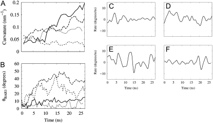
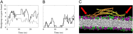
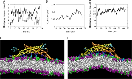
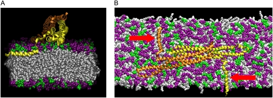
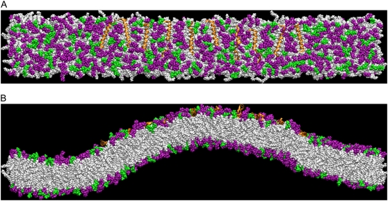
N-BAR domains are protein modules that bind to and induce curvature in membranes via a charged concave surface and N-terminal amphipathic helices. Recently, molecular dynamics simulations have demonstrated that the N-BAR domain can induce a strong local curvature that matches the curvature of the BAR domain surface facing the bilayer. Here we present further molecular dynamics simulations that examine in greater detail the roles of the concave surface and amphipathic helices in driving local membrane curvature. We find that the strong curvature induction observed in our previous simulations requires the stable presentation of the charged concave surface to the membrane and is not driven by the membrane-embedded amphipathic helices. Nevertheless, without these amphipathic helices embedded in the membrane, the N-BAR domain does not maintain a close association with the bilayer, and fails to drive membrane curvature. Increasing the membrane negative charge through the addition of PIP(2) facilitates closer association with the membrane in the absence of embedded helices. At sufficiently high concentrations, amphipathic helices embedded in the membrane drive membrane curvature independently of the BAR domain.


Figures

FIGURE 1
FIGURE 1
Membrane curvature and BAR domain orientation on the membrane surface. (A) Membrane curvature development for NBR1 (solid), NBR2 (long dash), NBR3 (short dash), and NBR− (dot). Simulations NBR1 and NBR2 were reported previously (20) and are presented here for comparison. Curvature is calculated as reported previously, except that only the thin bilayer section directly underneath the BAR domain is included, as opposed to the entire width along the short membrane axis (y axis). Each point is an average over 1 ns, with samples taken every 50 ps. (B) Altitudinal angle θBAR1 between the shortest principal axis of the BAR domain (initially directed along the y axis of the simulation cell) and the x, y plane (the plane of the membrane). Rate of change of the angle θBAR1 calculated from a smooth fit to the data in B for BAR domains in simulations NBR1 (C), NBR2 (D), NBR3 (E), and NBR− (F).
FIGURE 2
FIGURE 2
Binding of flexible BAR domain loops to lipid headgroups. The binding of Arg and Lys residues to oxygen atoms on lipid headgroups is shown for simulations NBR1 (A) and NBR3 (B). The solid and dashed lines represent the left and right loops, respectively, as depicted in the simulation snapshots (e.g., C). For this study, these loops comprise residues 161–171, inclusive. Residues are considered bound if the nitrogen and oxygen atoms remain within 4.2 Å for at least 1 ns (50-ps sampling interval). The binding of these loops assists in stabilizing the interaction of the BAR domain with the lipid bilayer (compare with Fig. 1, C and E). (C) Close-up snapshot showing the charged binding loops (red arrows) on either end of the BAR domain dissociating from the bilayer surface after 13 ns in simulation NBR3. The DOPC headgroups are purple and the DOPS headgroups are green. The lipid tails are white. Water and NaCl are also present in the simulations, but are left out of the image for clarity. In this and other close-up snapshots, only about half of the 45-nm membrane is shown.
FIGURE 3
FIGURE 3
Membrane interaction of the N-BAR with unbound N-terminal regions. (A) Binding of the charged end loops of the N-BAR domain to the DOPC/DOPS/PIP2 membrane in simulation NBRHP1. (B) Local curvature of the bilayer in the region of the N-BAR domain. (C) The binding of Arg and Lys residues on the BAR concave surface to oxygen atoms on the lipid headgroups. (D) Simulation snapshot at the beginning of the simulation showing the N-BAR domain with unbound N-terminal coil regions (dark cyan) and the lipid bilayer containing PC (purple), PS (green), and PIP2 (light cyan) headgroups. The basic residues on the protein (dark blue) interact strongly with the PIP2 headgroups even when the bilayer is relatively flat. (E) Simulation snapshot at 55 ns during development of local membrane curvature. About half of the 45-nm membrane is shown.
FIGURE 4
FIGURE 4
Orientation of the N-terminal helix. (A) Cross section along the long axis of the membrane showing the N-terminal helix embedded at the junction between the lipid headgroups (purple and green) and lipid tails (white). (B) Top view of the N-BAR domain showing both of the embedded N-terminal helices (red arrows). About half of the 45-nm membrane is shown.
FIGURE 5
FIGURE 5
Membrane curvature driven by amphipathic helices. (A) Top view of the membrane containing 10 embedded amphipathic helices (orange) after 36 ns. (B) Snapshot from the 10-helix simulation after 36 ns, showing the amphipathic helices inducing curvature in the membrane.

References

    1. McMahon, H. T., and J. L. Gallop. 2005. Membrane curvature and mechanisms of dynamic cell membrane remodelling. Nature. 438:590–596. - PubMed
    1. Zimmerberg, J., and M. M. Kozlov. 2006. How proteins produce cellular membrane curvature. Nat. Rev. Mol. Cell Biol. 7:9–19. - PubMed
    1. McMahon, H. T., and I. G. Mills. 2004. COP and clathrin-coated vesicle budding: different pathways, common approaches. Curr. Opin. Cell Biol. 16:379–391. - PubMed
    1. Bi, X., R. A. Corpina, and J. Goldberg. 2002. Structure of the Sec23/24-Sar1 pre-budding complex of the COPII vesicle coat. Nature. 419:271–277. - PubMed
    1. Nossal, R. 2001. Energetics of clathrin basket assembly. Traffic. 2:138–147. - PubMed

Publication types

LinkOut - more resources