. 2008 Jul;95(2):503-9.
doi: 10.1529/biophysj.107.121855. Epub 2008 May 9.

Toward resolution of ambiguity for the unfolded state

Affiliations

Toward resolution of ambiguity for the unfolded state

Gregory Beaucage. Biophys J. 2008 Jul.

Abstract

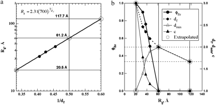
The unfolded states in proteins and nucleic acids remain weakly understood despite their importance in folding processes; misfolding diseases (Parkinson's and Alzheimer's); natively unfolded proteins (as many as 30% of eukaryotic proteins, according to Fink); and the study of ribozymes. Research has been hindered by the inability to quantify the residual (native) structure present in an unfolded protein or nucleic acid. Here, a scaling model is proposed to quantify the molar degree of folding and the unfolded state. The model takes a global view of protein structure and can be applied to a number of analytic methods and to simulations. Three examples are given of application to small-angle scattering from pressure-induced unfolding of SNase, from acid-unfolded cytochrome c, and from folding of Azoarcus ribozyme. These examples quantitatively show three characteristic unfolded states for proteins, the statistical nature of a protein folding pathway, and the relationship between extent of folding and chain size during folding for charge-driven folding in RNA.


Figures

FIGURE 1
FIGURE 1
Stylized model for protein structure. Schematic shows a rough two-dimensional cartoon of a protein chain, indicating the presence of a minimum path (gray circles) of p residues (circles) that differs from the protein's total residues (z) (gray and white circles) due to the presence of bridge/fold points. Fold points could be hydrophobic interactions, disulfide bonds, hydrogen bonds, or ionic bonds leading to association of the protein chain at large residue-index differences.
FIGURE 2
FIGURE 2
(a) Scattering from pH 5.5 100 bar SNase at 1% and 20°C (disklike regular object) (○) and 2800 bar, close to the unfolded state. Data is digitized from Panick et al. (29) and fit with the unified global function (solid lines) (18,23,25) that incorporates Eqs. 1. and 2, shown as dashed curves for the 2800-bar fit. (b) Fractal analysis of scattering for the transition from a PMG-like regular object (large rectangle) to a projected unfolded linear chain (large circle) at ∼4000 bar, where dmin = df and c = 1. (c) Minimum path, p; number of residues, z; and extent of folding, fBr versus pressure, predicting the same pressure range for the purely unfolded state, where p = z and fBr = 0. (Curves are empirical fits to 1 − k0exp(k1P2) and k0exp(k1P2).) (d) SNase in the native state (pH ∼7 atmospheric pressure) from PDB using King 1.39 viewer (28).
FIGURE 3
FIGURE 3
Unified fits (18,19,23,25) to x-ray scattering data from Caliskan et al. (39) and Chauchan et al. (40) on the 195-nucleotide Azoarcus ribozyme for variable Mg2+ concentrations. (a) 4.26 mM (N). (b) 0.59 mM (Ic). (c) 0.47 mM (U).
FIGURE 4
FIGURE 4
Scattering-fit results and extrapolated values for the data from Fig. 3 (39,40) on Azoarcus ribozyme for variable Mg2+ concentrations. (a) Rg versus 1/df. Limits of df are 3, 2, and 5/3 for three-dimensional, Gaussian, and good-solvent (self-avoiding walk) states, respectively. From this plot, the limiting sizes (circles) can be determined and used to extend the measured dimensions and φBr in b. (b) Mole fraction folding (left), φBr, and three measured dimensions, with the limiting values (right) showing decreasing tortuosity, dmin, and increasing connectivity, c, with folding (lower Rg). (Circled values at Rg = 20.5, 61.2, and 117.7 Å are predicted for df = 3, 2, and 5/3, respectively.) Horizontal dashed lines indicate df = 2 and 5/3.

References

    1. Pande, V. S., and D. S. Rokhsar. 1998. Is the molten globule a third phase of proteins? Proc. Natl. Acad. Sci. USA. 95:1490–1494. - PMC - PubMed
    1. Uversky, V. N. 2003. Protein folding revisited. A polypeptide chain at the folding-misfolding-nonfolding cross-roads: which way to go? Cell. Mol. Life Sci. 60:1852–1871. - PMC - PubMed
    1. Chaudhuri, T. K., and S. Paul. 2006. Protein-misfolding diseases and chaperone-based therapeutic approaches. FEBS J. 273:1331–1349. - PubMed
    1. Nissen, P., J. Hansen, N. Ban, P. B. Moore, and T. A. Steitz. 2000. The structural basis of ribosome activity in peptide bond synthesis. Science. 289:920–930. - PubMed
    1. Tanford, C. 1968. Protein denaturation. Adv. Protein Chem. 23:121–282. - PubMed

LinkOut - more resources