. 2008 Aug;95(4):1902-12.
doi: 10.1529/biophysj.108.129957. Epub 2008 May 9.

Balance between ultrafast parallel reactions in the green fluorescent protein has a structural origin

Affiliations

Balance between ultrafast parallel reactions in the green fluorescent protein has a structural origin

Jasper J van Thor et al. Biophys J. 2008 Aug.

Abstract

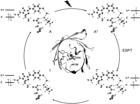
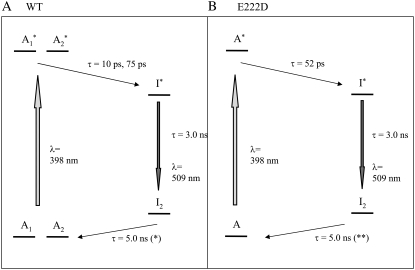
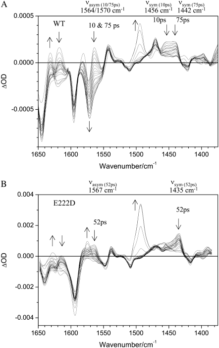
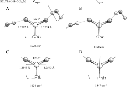
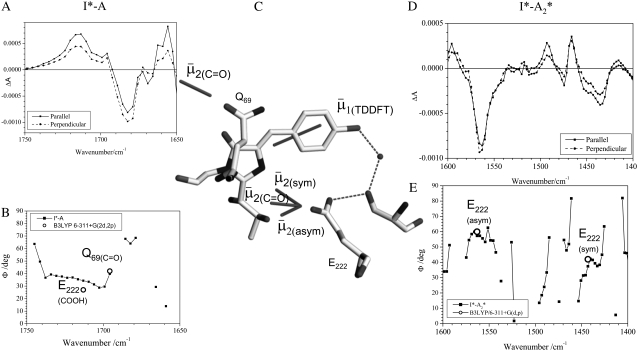
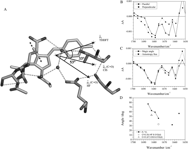
The fluorescence photocycle of the green fluorescent protein is functionally dependent on the specific structural protein environment. A direct relationship between equilibrium protein side-chain conformation of glutamate 222 and reactivity is established, particularly the rate of ultrafast proton transfer reactions in the fluorescence photocycle. We show that parallel transformations in the photocycle have a structural origin, and we report on the vibrational properties of responsive amino acids on an ultrafast timescale. Blue excitation of GFP drives two parallel, excited-state deuteron transfer reactions with 10 ps and 75 ps time constants to the buried carboxylic acid side chain of glutamate 222 via a hydrogen-bonding network. Assignment of 1456 cm(-1) and 1441 cm(-1) modes to nu(sym) and assignment of 1564 cm(-1) and 1570 cm(-1) features to nu(asym) of E222 in the 10 ps and 75 ps components, respectively, was possible from the analysis of the transient absorption data of an E222D mutant and was consistent with photoselection measurements. In contrast to the wild-type, measurements of E222D can be described with only one difference spectrum, with the nu(sym) mode at 1435 cm(-1) and the nu(asym) mode at 1567 cm(-1), also correlating a large Deltanu(asym-sym) with slow excited-state proton transfer kinetics. Density Functional Theory calculations and published model compound and theoretical studies relate differences in Deltanu(asym-sym) to the strength and number of hydrogen-bonding interactions that are detected via equilibrium geometry and COO- stretching frequency differences of the carboxylate. The correlation of photocycle kinetics with side-chain conformation of the acceptor suggests that proton transfer from S205 to E222 controls the rate of the overall excited-state proton transfer process, which is consistent with recent theoretical predictions. Photoselection measurements show agreement for localized C=O vibrations of chromophore, Q69, and E222 with Density Functional Theory and ab initio calculations placed in the x-ray geometry and provide their vibrational response in the intermediates in the photocycle.


Figures

FIGURE 1
FIGURE 1
Overview of structural, H-bonding, and electronic changes in the fluorescence photocycle. The phenolic oxygen of the HBDI chromophore is H-bonded indirectly to glutamate 222 via a water molecule and serine 205. Coordinates are taken from PDB 1W7S (5). The electronically excited singlet state of the neutral and anion chromophore, A* and I* respectively, are indicated. ESPT reactions occur during the transition between the A* and I* states that have 10 and 75 ps time constant in D2O (4,10). Blue excitation with 400 nm light is indicated to create the A* state.
FIGURE 2
FIGURE 2
Photocycle scheme for the (A) wild-type and the (B) E222D mutant used for global fitting of the transient absorption data. Note that the I1 intermediate is not considered here because it does not accumulate to an observable level in the photocycle and is only significant in the pump-dump-probe experiments outlined in reference 10.
FIGURE 3
FIGURE 3
Transient difference absorption changes in the mid-infrared with excitation at 400 nm for (A) wild-type and (B) mutant E222D. (A) Difference absorption relative to the ground state between 1650 cm−1 and 1375 cm−1 with delays between 2 ps and 2 ns of wild-type GFP. Arrows indicate changes with time. The frequencies of the νasym and νsym modes of E222 are indicated for the 10 ps and 75 ps components for the excited-state deuteron transfer reactions. (B) Difference absorption data with the same delays shown for the E222D mutant. The νasym and νsym modes of D222 are found at discrete frequencies for the excited state deuteron transfer reaction, which has a 52 ps time constant.
FIGURE 4
FIGURE 4
DFT optimized geometry and frequencies for νasym and νsym normal modes for isolated and interacting E222 at the B3LYP 6-311+G(2p,2d) level. (A) Bond angle and lengths for a geometry optimized model including hydrogen-bonding interactions to water Z223 and S205. Displacement vectors and calculated frequency of νasym are shown. (B) Displacement vectors and frequency of νsym for interacting E222. (C) Bond angle and lengths for a geometry optimized model excluding hydrogen-bonding interactions but maintaining dihedral angles taken from the x-ray geometry, and vectors and calculated frequency of νasym. (D) Displacement vectors and frequency of νsym for isolated E222
FIGURE 5
FIGURE 5
Correspondence of theoretical transition dipole moments of assigned amino acid normal modes with polarization-dependent species associated difference spectra. (A) The 1750–1650 cm−1 region of the polarization dependent I* minus A difference spectrum of the wild-type. COOH and C=O stretching vibrations of E222 and Q69 are assigned at 1711 and 1695 cm−1, respectively. (B) Calculated and experimental angles formula image in the 1750–1650 cm−1 region of the polarization-dependent I* minus A difference spectrum. (C) Structural arrangement determined in the ground state with coordinates taken from PDB 1W7S (4). The optical transition dipole moment formula image obtained from a TD-DFT B3LYP/6-311+G(d,p) calculation is shown as an in-plane vector. The vibrational transition dipole moment vectors formula image from finite difference calculations at the B3LYP/6-311+G(d,p) are placed in the x-ray geometry. (D) The 1600–1400 cm−1 region of the polarization dependent I* minus A2* difference spectrum of the wild-type. Theoretical and experimental angles formula image in the 1600–1400 cm−1 region of the polarization-dependent I* minus A2* difference spectrum.
FIGURE 6
FIGURE 6
Reorientation of the chromophore C=O vibrational transition dipole moment in the excited state. (A) Orientation and angles between the theoretical optical transition dipole moments formula image and formula image for the chromophore C=O vibration in the ground and excited state at the HF/6-31G(d) and CIS/6-31G(d) level, respectively. The general direction of the normal mode displacement is shown with arrows. (B) The 1700–1650 cm−1 region of the polarization dependent A1* minus A difference spectrum of the wild-type. (C) Reconstructed anisotropy-free ((A+ 2A)/3) A1* minus A species-associated difference spectrum and that from measurements at magic angle show good agreement. (D) Theoretical and experimental angles formula image in the 1700–1650 cm−1 region of the A1* minus A difference spectrum.

References

    1. Tsien, R. Y. 1998. The green fluorescent protein. Annu. Rev. Biochem. 67:509–544. - PubMed
    1. Weber, W., V. Helms, J. A. McCammon, and P. W. Langhoff. 1999. Shedding light on the dark and weakly fluorescent states of green fluorescent proteins. Proc. Natl. Acad. Sci. USA. 96:6177–6182. - PMC - PubMed
    1. Usman, A., O. F. Mohammed, E. T. Nibbering, J. Dong, K. M. Solntsev, and L. M. Tolbert. 2005. Excited-state structure determination of the green fluorescent protein chromophore. J. Am. Chem. Soc. 127:11214–11215. - PubMed
    1. van Thor, J. J., G. Zanetti, K. Ronayne, and M. Towrie. 2005. Structural events in the photocycle of green fluorescent protein. J. Phys. Chem. B. 109:16099–16108. - PubMed
    1. van Thor, J. J., G. Y. Georgiev, M. Towrie, and J. T. Sage. 2005. Ultrafast and low barrier motions in the photoreactions of the green fluorescent protein. J. Biol. Chem. 280:33652–33659. - PubMed

Publication types

MeSH terms

Substances