. 2008 Jul;7(7):1158-67.
doi: 10.1128/EC.00434-07. Epub 2008 May 9.

Direct interactions between the Paf1 complex and a cleavage and polyadenylation factor are revealed by dissociation of Paf1 from RNA polymerase II

Affiliations

Direct interactions between the Paf1 complex and a cleavage and polyadenylation factor are revealed by dissociation of Paf1 from RNA polymerase II

Kristen Nordick et al. Eukaryot Cell. 2008 Jul.

Abstract

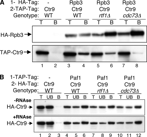
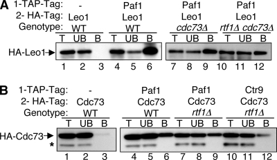
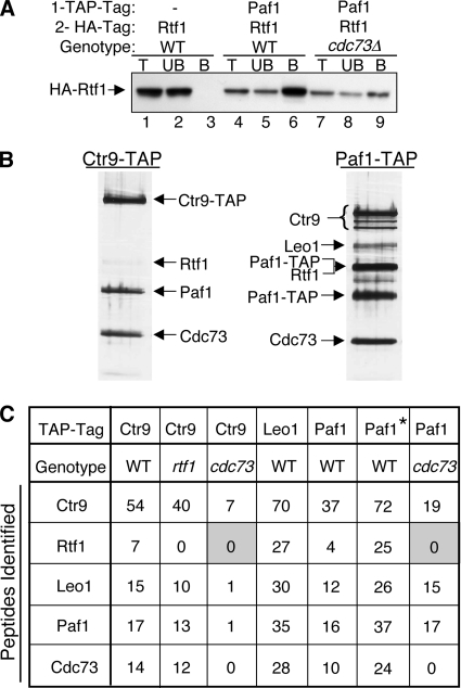
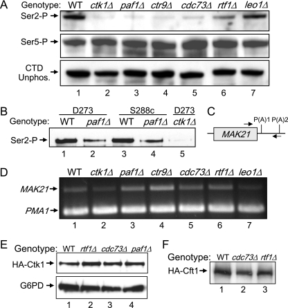
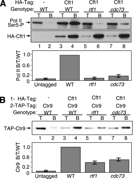
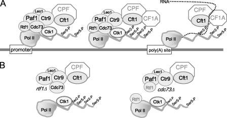
The Paf1 complex (Paf1, Ctr9, Cdc73, Rtf1, and Leo1) is normally associated with RNA polymerase II (Pol II) throughout the transcription cycle. However, the loss of either Rtf1 or Cdc73 results in the detachment of the Paf1 complex from Pol II and the chromatin form of actively transcribed genes. Using functionally tagged forms of the Paf1 complex factors, we have determined that, except for the more loosely associated Rtf1, the remaining components stay stably associated with one another in an RNase-resistant complex after dissociation from Pol II and chromatin. The loss of Paf1, Ctr9, or to a lesser extent Cdc73 or Rtf1 results in reduced levels of serine 2 phosphorylation of the Pol II C-terminal domain and in increased read through of the MAK21 polyadenylation site. We found that the cleavage and polyadenylation factor Cft1 requires the Pol II-associated form of the Paf1 complex for full levels of interaction with the serine 5-phosphorylated form of Pol II. When the Paf1 complex is dissociated from Pol II, a direct interaction between Cft1 and the Paf1 complex can be detected. These results are consistent with the Paf1 complex providing a point of contact for recruitment of 3'-end processing factors at an early point in the transcription cycle. The lack of this connection helps to explain the defects in 3'-end formation observed in the absence of Paf1.


Figures

FIG. 1.
FIG. 1.
The mutation of RTF1 or CDC73 results in the dissociation of Ctr9 from Pol II but not from Paf1. (A) The loss of Rtf1 or Cdc73 reduces the co-IP of TAP-tagged Ctr9 with HA-tagged Rpb3. Protein extracts from strains bearing the indicated combinations of tags and deletion mutations were subjected to IP using an α-HA antibody as described in Materials and Methods. Ten-microgram samples of total protein (T) and the bound pellets (B) were probed for the immunoprecipitated HA-tagged Rpb3 (upper panel) and the coimmunoprecipitated TAP-tagged Ctr9 (lower panel). The strains used were YJJ1329 (lanes 1 and 2), YJJ1857 (lanes 3 and 4), YJJ1859 (lanes 5 and 6), and YJJ1861 (lanes 7 and 8). − represents the absence of protein with the indicated tag. (B) The association of Paf1 with Ctr9 is not affected by dissociation from Pol II or by RNase treatment. TAP-tagged Paf1 was immunoprecipitated, and the presence of coimmunoprecipitated HA-tagged Ctr9 was detected in samples incubated without (−RNAse; upper panel) or with (+RNAse; lower panel) RNase A treatment prior to the IP. Ten-microgram samples of total protein (T), an equal volume of the unbound IP supernatant (UB), and the bound pellets (B) were probed as indicated. The strains used were YJJ1599 (lanes 1 to 3), YJJ1782 (lanes 4 to 6), YJJ1784 (lanes 7 to 9), and YJJ1803 (lanes 10 to 12).
FIG. 2.
FIG. 2.
There is still an association of Leo1 and Cdc73 with Paf1 and Ctr9 after the Paf1C is dissociated from Pol II. Protein extracts from strains bearing the indicated combinations of tags and mutations were subjected to IP using an α-TAP antibody, and coimmunoprecipitates were detected with an α-HA antibody as described in Materials and Methods. Lane labeling is as described in the legend to Fig. 1B. − represents the absence of protein with the indicated tag. (A) HA-tagged Leo1 coimmunoprecipitates with TAP-tagged Paf1 in the absence of Cdc73 or Rtf1. The strains used were YJJ1330 (lanes 1 to 3), YJJ1786 (lanes 4 to 6), YJJ1787 (lanes 7 to 9), and YJJ1809 (lanes 10 to 12). (B) HA-tagged Cdc73 coimmunoprecipitates with TAP-tagged Paf1 and Ctr9 in the absence of Rtf1. The strains used were YJJ1583 (lanes 1 to 3), YJJ1832 (lanes 4 to 6), YJJ1834 (lanes 7 to 9), and YJJ1841 (lanes 10 to 12). The asterisk denotes an HA-cross-reacting band.
FIG. 3.
FIG. 3.
The association of Rtf1 with the Paf1C is reduced in the absence of Cdc73. (A) TAP-tagged Paf1 was immunoprecipitated, and the presence of coimmunoprecipitated HA-tagged Rtf1 was detected. The strains used were YJJ1589 (lanes 1 to 3), YJJ1789 (lanes 4 to 6), and YJJ1791 (lanes 7 to 9). Lane labeling is as described in the legend to Fig. 1B. − indicates the absence of TAP-tagged Paf1. (B) Large-scale purification of TAP-tagged Ctr9 (YJJ1857) and Paf1 (YJJ1782) from WT cells was performed as described in Materials and Methods. Copurifying proteins were separated by gel electrophoresis, silver stained, and identified by mass spectrometry as indicated. (C) Proteins associated with TAP-tagged Ctr9, Leo1, and Paf1 in strains with the indicated genetic backgrounds were analyzed by mass spectrometry as described in Materials and Methods. The numbers of identified peptides of the indicated Paf1C components are listed. The asterisk marking the second column corresponding to Paf1-TAP and the WT genotype indicates a preparation done using a buffer with a lower salt concentration (150 mM NaCl) than that in the other samples. The lower-molecular-weight Paf1-TAP band was the result of the proteolytic degradation of the amino terminus. Shading highlights the lack of detection of Rtf1 peptides. The strains used were Ctr9-TAP-expressing WT YJJ1873, Ctr9-TAP-expressing rtf1 mutant YJJ1875, Ctr9-TAP-expressing cdc73 mutant YJJ1861, Leo1-TAP-expressing WT YN96, Paf1-TAP-expressing WT YJJ1782, and Paf1-TAP-expressing cdc73 mutant YJJ1787.
FIG. 4.
FIG. 4.
(A) The loss of some Paf1C components reduces Pol II CTD Ser2-P. Protein extracts prepared from isogenic strains bearing the indicated mutations were separated by electrophoresis, blotted, and probed for the indicated antigens as described in Materials and Methods. The strains used were YJJ662 (WT), YJJ1821 (ctk1Δ), YJJ577 (paf1Δ), YJJ1197 (ctr9Δ), YJJ665 (cdc73Δ), YJJ1303 (rtf1Δ), and YJJ1336 (leo1Δ). Antibodies used for Pol II were H5 (α-Ser2-P), H14 (α-Ser5-P), and 8WG16 (α-unphosphorylated CTD [CTD unphos.]). (B) The reduction of Pol II CTD Ser2-P by the loss of Paf1 is not yeast strain dependent. Protein extracts from pairs of isogenic strains with the indicated genetic backgrounds were analyzed for the Pol II Ser2-P CTD as described in the legend to panel A by using the BL2894 antibody. The strains used were YJJ662 (WT; lane 1), YJJ664 (paf1Δ; lane 2), YSB2116 (WT; lane 3), YSB2118 (paf1Δ; lane 4), and YJJ1871 (ctk1Δ; lane 5). (C) Schematic of the MAK21 gene showing the locations of primers spanning the first poly(A) site [P(A)1] used for RT-PCRs. (D) The loss of some Paf1C factors, but not Leo1 or Ctk1, increases the read through of the MAK21 poly(A) site. RT-PCR was used to measure the amount of the MAK21 read-through transcript, which was normalized to the signal from the PMA1 gene. Strains used for RNA isolation were the same as those listed in the legend to panel A. (E) The loss of Rtf1 or Cdc73 does not affect the abundance of HA-tagged Ctk1. Protein extracts from isogenic HA-tagged Ctk1 strains were analyzed as described in the legend to panel A. The strains used were YJJ1873 (WT), YJJ1875 (rtf1Δ), YJJ1897 (cdc73Δ), and YJJ1882 (paf1Δ). Extracts in the upper panel were probed with α-HA. Those in the lower panel were probed with α-G6PD. (F) The loss of Rtf1 or Cdc73 does not affect the abundance of HA-tagged Cft1. Protein extracts from isogenic HA-tagged Cft1 strains were analyzed as described in the legend to panel A and probed with α-HA. The strains used were YJJ1864 (WT), YJJ1850 (cdc73Δ), and YJJ1849 (rtf1Δ).
FIG. 5.
FIG. 5.
Dissociation of the Paf1C from Pol II reduces the association of polyadenylation factor Cft1 with Pol II and reveals a direct association between the Paf1C and Cft1. Extracts from strains bearing the indicated tagged proteins and mutations were immunoprecipitated with α-HA antibody and then probed with appropriate antibodies. Signals in the lower panels were quantitated as described in Materials and Methods as the ratio of Pol II bound to total protein-, and the results are expressed relative to the WT ratio, which was set at 1. T, 10 μg of total protein; B, bound IP pellet; −, absence of HA-tagged Cft1. (A) The loss of Rtf1 or Cdc73 reduces the association of Cft1 with Pol II. HA-tagged Cft1 was immunoprecipitated, and the presence of the Ser5-P CTD form of Pol II was detected with the α-Ser5-P CTD H14 antibody (upper panel). The protein blot was stripped and reprobed with α-HA for the presence of HA-tagged Cft1 (middle panel). The error bars in the graph in the lower panel represent the standard deviations of data from four repetitions of the experiment. The strains used were YJJ1824 (lanes 1 and 2), YJJ1864 (lanes 3 and 4), YJJ1849 (lanes 5 and 6), and YJJ1850 (lanes 7 and 8). (B) The Paf1C and Cft1 interact in the absence of Rtf1 or Cdc73. HA-tagged Cft1 was immunoprecipitated, and the presence of coimmunoprecipitated TAP-tagged Ctr9 was detected with α-TAP (upper panel). The solid bars representing quantitated data in the lower panel indicate the averages and the error bars show the ranges of results obtained from replicate experiments. The strains used were YJJ1824 (lanes 1 and 2), YJJ1864 (lanes 3 and 4), YJJ1849 (lanes 5 and 6), and YJJ1850 (lanes 7 and 8).
FIG. 6.
FIG. 6.
The Paf1C provides one of several contacts to recruit polyadenylation factors to elongating Pol II. Shown is a simplified model describing the interactions elucidated in this work within the Paf1C and between the Paf1C and Ctk1 and Cft1 in WT, rtf1Δ, and cdc73Δ strains as described in Discussion.

Similar articles

Cited by

References

    1. Ahn, S. H., M. Kim, and S. Buratowski. 2004. Phosphorylation of serine 2 within the RNA polymerase II C-terminal domain couples transcription and 3′ end processing. Mol. Cell 1367-76. - PubMed
    1. Akanuma, T., S. Koshida, A. Kawamura, Y. Kishimoto, and S. Takada. 2007. Paf1 complex homologues are required for Notch-regulated transcription during somite segmentation. EMBO Rep. 8858-863. - PMC - PubMed
    1. Barilla, D., B. A. Lee, and N. J. Proudfoot. 2001. Cleavage/polyadenylation factor IA associates with the carboxyl-terminal domain of RNA polymerase II in Saccharomyces cerevisiae. Proc. Natl. Acad. Sci. USA 98445-450. - PMC - PubMed
    1. Bentley, D. L. 2005. Rules of engagement: co-transcriptional recruitment of pre-mRNA processing factors. Curr. Opin. Cell Biol. 17251-256. - PubMed
    1. Betz, J. L., M. Chang, T. M. Washburn, S. E. Porter, C. L. Mueller, and J. A. Jaehning. 2002. Phenotypic analysis of Paf1/RNA polymerase II complex mutations reveals connections to cell cycle regulation, protein synthesis, and lipid and nucleic acid metabolism. Mol. Genet. Genomics 268272-285. - PubMed

Publication types

MeSH terms

Substances