

Identification and characterization of a novel Mdm2 splice variant acutely induced by the chemotherapeutic agents adriamycin and actinomycin D
- PMID: 18469520
- PMCID: PMC3608406
- DOI: 10.4161/cc.7.11.5985
Identification and characterization of a novel Mdm2 splice variant acutely induced by the chemotherapeutic agents adriamycin and actinomycin D
Abstract
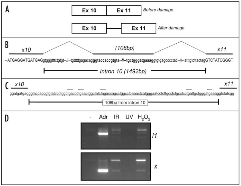
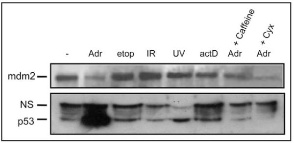
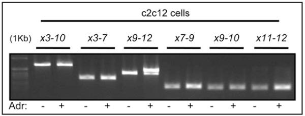
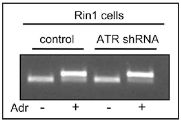
Mdm2, as the most important negative regulator of p53, plays an important homeostatic role in regulating cell division and the cellular response to DNA damage, oncogenic insult and other forms of cellular stress. We discovered that the DNA damaging agent adriamycin (doxorubicin) induces a novel aberrantly spliced Mdm2 mRNA which incorporates 108 bp of intronic sequence not normally found in the Mdm2 mature mRNA. Accordingly, we term this Mdm2 splice variant Mdm2(+108). Importantly, this insertion introduces in-frame nonsense codons, thus encoding a profoundly truncated mdm2 protein lacking the C-terminal RING finger domain and the E3 ubiquitin ligase activity. A wide range of pharmacological testing revealed that Mdm2(+108) is induced, in mouse and rat cells, in specific response to Adriamycin and actinomycin D, but not other modes of DNA damage. Meanwhile, antibodies against the N-terminal region of mdm2 reveal a marked reduction in detectable mdm2 protein upon Adriamycin treatment, while p53 accumulates to strikingly high levels. We thus conclude that this alternative spicing of Mdm2 may be an important mechanism to facilitate massive accumulation of p53 in response to genotoxic agents.
Figures
Similar articles
-
[Any way you splice it: Mdm2 at the crossroads of tumor surveillance].Ai Zheng. 2008 Sep;27(9):993-7. Ai Zheng. 2008. PMID: 18799043 Review. Chinese.
-
Mdm2 Splice isoforms regulate the p53/Mdm2/Mdm4 regulatory circuit via RING domain-mediated ubiquitination of p53 and Mdm4.Cell Cycle. 2017 Apr 3;16(7):660-664. doi: 10.1080/15384101.2017.1288327. Epub 2017 Feb 6. Cell Cycle. 2017. PMID: 28166445 Free PMC article.
-
Dissecting the p53-Mdm2 feedback loop in vivo: uncoupling the role in p53 stability and activity.Oncotarget. 2014 Mar 15;5(5):1149-56. doi: 10.18632/oncotarget.1797. Oncotarget. 2014. PMID: 24658419 Free PMC article.
-
MDM2 does not influence p53-mediated sensitivity to DNA-damaging drugs.Mol Cancer Ther. 2002 Oct;1(12):1097-104. Mol Cancer Ther. 2002. PMID: 12481433
-
Unlocking the Mdm2-p53 loop: ubiquitin is the key.Cell Cycle. 2008 Feb 1;7(3):287-92. doi: 10.4161/cc.7.3.5358. Epub 2007 Nov 25. Cell Cycle. 2008. PMID: 18235222 Review.
Cited by
-
The MDM2-a splice variant of MDM2 alters transformation in vitro and the tumor spectrum in both Arf- and p53-null models of tumorigenesis.Mol Cancer Res. 2009 Jun;7(6):863-9. doi: 10.1158/1541-7786.MCR-08-0418. Epub 2009 Jun 2. Mol Cancer Res. 2009. PMID: 19491200 Free PMC article.
-
Genotoxic stress modulates CDC25C phosphatase alternative splicing in human breast cancer cell lines.Mol Oncol. 2012 Oct;6(5):542-52. doi: 10.1016/j.molonc.2012.06.003. Epub 2012 Jul 27. Mol Oncol. 2012. PMID: 22871320 Free PMC article.
References
-
- Vogelstein B, Kinzler KW. Cancer genes and the pathways they control. Nature medicine. 2004;10:789–99. - PubMed
-
- Cordon-Cardo C, Latres E, Drobnjak M, Oliva MR, Pollack D, Woodruff JM, Marechal V, Chen J, Brennan MF, Levine AJ. Molecular abnormalities of mdm2 and p53 genes in adult soft tissue sarcomas. Cancer research. 1994;54:794–9. - PubMed
-
- Bond GL, Hu W, Levine AJ. MDM2 is a central node in the p53 pathway: 12 years and counting. Current cancer drug targets. 2005;5:3–8. - PubMed
Publication types
MeSH terms
Substances
Grants and funding
LinkOut - more resources
Full Text Sources
Molecular Biology Databases
Research Materials
Miscellaneous