. 2008 Jun 26;453(7199):1253-7.
doi: 10.1038/nature06963. Epub 2008 May 11.

Rapid strengthening of thalamo-amygdala synapses mediates cue-reward learning

Affiliations

Rapid strengthening of thalamo-amygdala synapses mediates cue-reward learning

Kay M Tye et al. Nature. .

Abstract

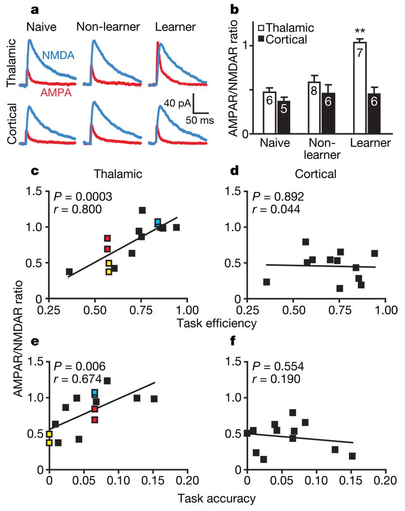
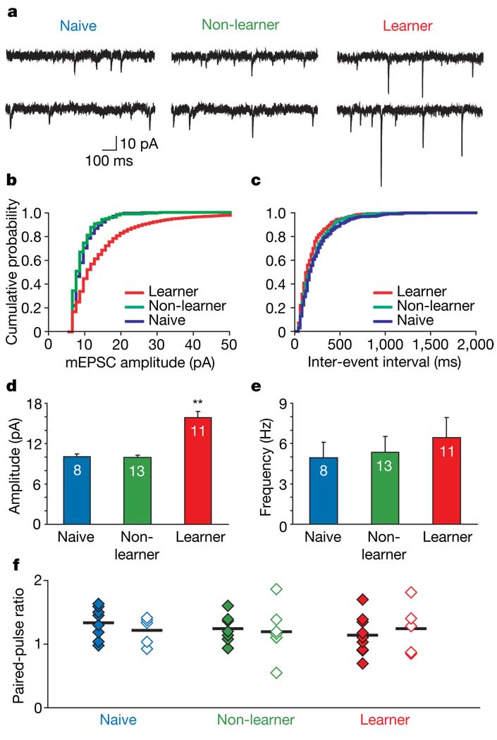
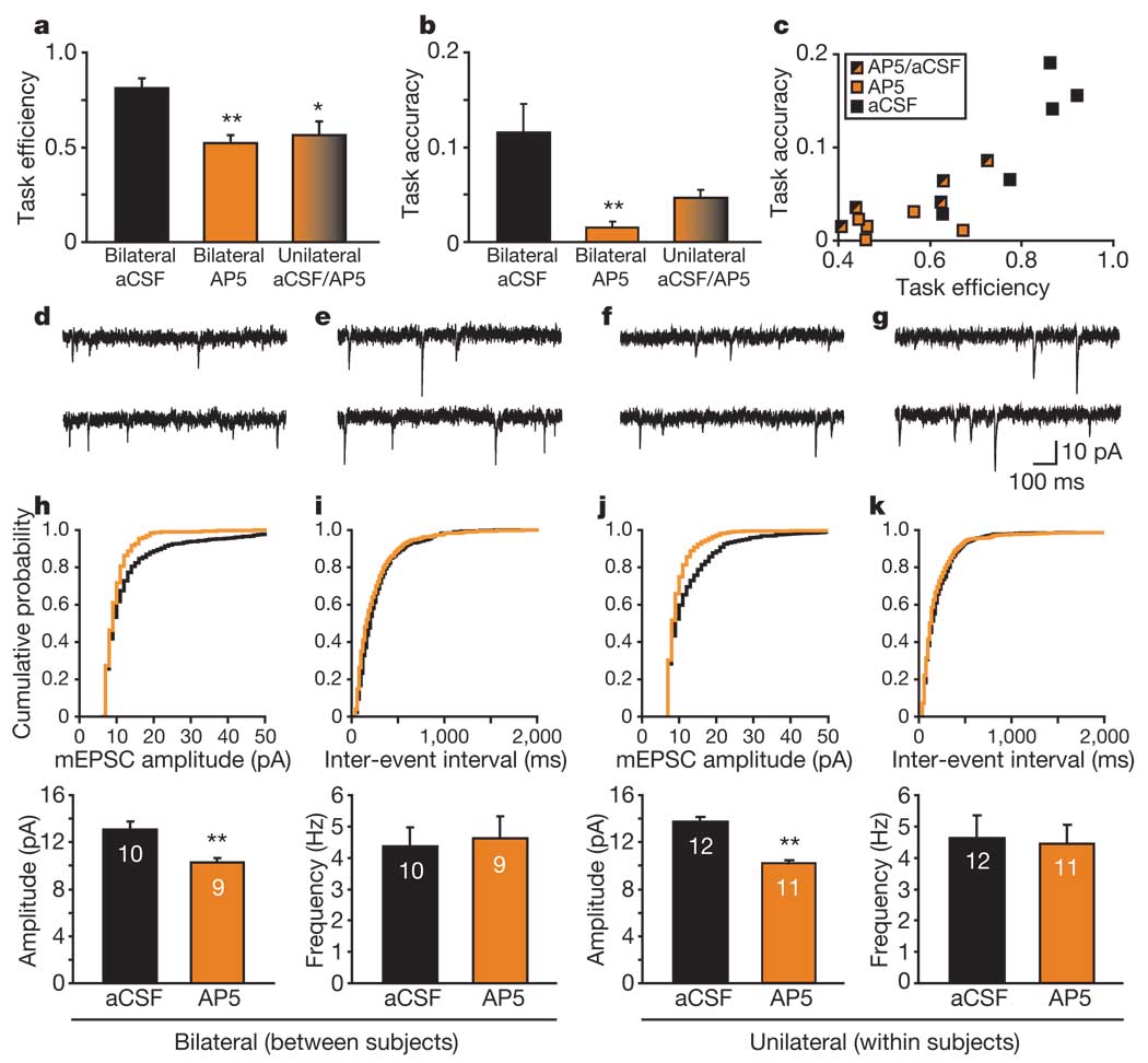
What neural changes underlie individual differences in goal-directed learning? The lateral amygdala (LA) is important for assigning emotional and motivational significance to discrete environmental cues, including those that signal rewarding events. Recognizing that a cue predicts a reward enhances an animal's ability to acquire that reward; however, the cellular and synaptic mechanisms that underlie cue-reward learning are unclear. Here we show that marked changes in both cue-induced neuronal firing and input-specific synaptic strength occur with the successful acquisition of a cue-reward association within a single training session. We performed both in vivo and ex vivo electrophysiological recordings in the LA of rats trained to self-administer sucrose. We observed that reward-learning success increased in proportion to the number of amygdala neurons that responded phasically to a reward-predictive cue. Furthermore, cue-reward learning induced an AMPA (alpha-amino-3-hydroxy-5-methyl-isoxazole propionic acid)-receptor-mediated increase in the strength of thalamic, but not cortical, synapses in the LA that was apparent immediately after the first training session. The level of learning attained by individual subjects was highly correlated with the degree of synaptic strength enhancement. Importantly, intra-LA NMDA (N-methyl-d-aspartate)-receptor blockade impaired reward-learning performance and attenuated the associated increase in synaptic strength. These findings provide evidence of a connection between LA synaptic plasticity and cue-reward learning, potentially representing a key mechanism underlying goal-directed behaviour.


Figures

Figure 1
Figure 1. Reward-related learning success is correlated with rapid increases in cue-related firing
a, Diagram of operant chamber from above. b, Schematic of behavioural paradigm. c, Temporal dynamics of neuronal population response to the reward-predictive cue. Spike activity of all simultaneously recorded LA units (n = 13) from a rat that successfully acquired the task during the first session; 100-ms bins. This population of neurons develops a response to the onset of the reward-predictive cue with task acquisition (acq.). d, Peri-event histogram of a single LA neuron from a different rat that successfully acquired the task in the first session; 29 trials in each epoch. e, Population histograms (50-ms bins, error bars indicate s.e.m.) of the mean Z-score for all neurons recorded during session 1 (grey; n = 95 neurons) and session 3 (red; n = 123 neurons) for all rats (n = 7). Two asterisks, P < 0.004. f, g, Correlation between proportion of cue-responsive neurons and (f) task efficiency and (g) task accuracy across three sessions. Colours indicate the same rat on different sessions. Only rats with at least six neurons per session were included in scatter plots (n = 6). For all peri-event histograms, time zero indicates cue onset.
Figure 2
Figure 2. Degree of AMPAR/NMDAR enhancement predicts cue–reward learning
a, EPSCs evoked by stimulation of thalamic or cortical afferents in rats that were naive, non-learners or learners. b, AMPAR/NMDAR ratios evoked from thalamic afferents were significantly increased in learners (n = 6 rats) in comparison with non-learners (n = 6 rats) or naives (n = 5 rats). Numbers in bars indicate numbers of cells; error bars indicate s.e.m. Two asterisks, P < 0.001, significant difference from other groups as well as from cortical afferent. c–f, Correlation between AMPAR/NMDAR ratio and either task efficiency (c, d) or task accuracy (e, f) for EPSCs evoked from thalamic (c, e), but not cortical (d, f), pathways; the subjects were the same as in b. Colours indicate multiple cells recorded from the same rat; black indicates single cells recorded from each rat.
Figure 3
Figure 3. Successful cue–reward learning induces an increase in mEPSC amplitude but not in frequency or paired-pulse ratio
a, Sample mEPSCs from rats that were naive, non-learners or learners. b, c, Cumulative probability plots of amplitude (b) and frequency (c) for representative neurons from each group; bins were 1 pA (b) and 20 ms (c). d, e, Learners (n = 6 rats) had increased mEPSC amplitude (d), but not frequency (e), relative to non-learners (n = 6 rats) and naive rats (n = 5 rats). Numbers in bars indicate numbers of cells; errors bars indicate s.e.m. Two asterisks, P < 0.001. f, Lack of change in paired-pulse ratio between the different groups of rats. Filled diamonds, ratios evoked from the thalamic pathway; open diamonds, ratios evoked from the cortical pathway; horizontal lines indicate the means.
Figure 4
Figure 4. Local NMDAR blockade attenuates reward-related learning and the associated increase in mEPSC amplitude
a, b, Measures of task performance among groups (n = 5 rats per group). Task efficiency (a) is decreased after either unilateral or bilateral AP5 intra-LA infusion, whereas task accuracy (b) is decreased after bilateral AP5 (asterisk, P < 0.05, two asterisks, P < 0.009, compared with aCSF). No other comparisons were significant. Error bars indicate s.e.m. c, Individual rat performances based on task efficiency and task accuracy. d–g, Sample mEPSCs from rats that received pre-training infusions. d, e, Traces recorded from rats that received bilateral infusions of AP5 (d) and aCSF (e). f, g, Traces recorded from a representative rat that received unilateral intra-LA infusions of AP5 (f) and aCSF (g). h–k, Cumulative probability plots of amplitude (h, j) and frequency (i, k) for mEPSCs in representative cells from rats receiving bilateral infusions of AP5 (orange) or aCSF (black) (h, i) or unilateral infusions of AP5 and aCSF (j, k). Below each probability plot is the corresponding bar graph indicating the group mean and s.e.m. There is a difference in amplitude (h, j), but not in frequency (i, k) for both bilaterally infused (h) and unilaterally infused (j) rats; two asterisks, P < 0.001, compared with aCSF. Numbers in bars indicate numbers of cells.

References

    1. LeDoux J. The emotional brain, fear, and the amygdala. Cell. Mol. Neurobiol. 2003;23:727–738. - PMC - PubMed
    1. Davis M. In: The Amygdala: Neurobiological Aspects of Emotion, Memory, and Mental Dysfunction. Aggleton JP, editor. Chichester, UK: Wiley; 1992. pp. 255–306.
    1. Rosenkranz JA, Grace AA. Dopamine-mediated modulation of odour-evoked amygdala potentials during pavlovian conditioning. Nature. 2002;417:282–287. - PubMed
    1. Maren S, Quirk GJ. Neuronal signalling of fear memory. Nature Rev. Neurosci. 2004;5:844–852. - PubMed
    1. Cador M, Robbins TW, Everitt BJ. Involvement of the amygdala in stimulus-reward associations: interaction with the ventral striatum. Neuroscience. 1989;30:77–86. - PubMed

Publication types