Comparative Study
. 2008 Jun;40(6):776-81.
doi: 10.1038/ng.149. Epub 2008 May 11.

X-linked protocadherin 19 mutations cause female-limited epilepsy and cognitive impairment

Affiliations
Comparative Study

X-linked protocadherin 19 mutations cause female-limited epilepsy and cognitive impairment

Leanne M Dibbens et al. Nat Genet. 2008 Jun.

Abstract

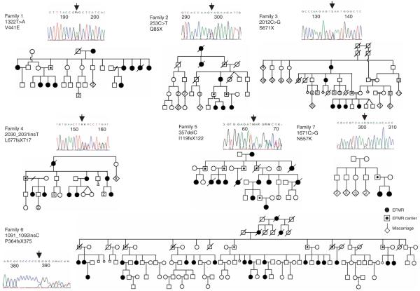
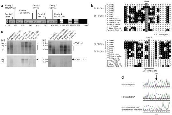
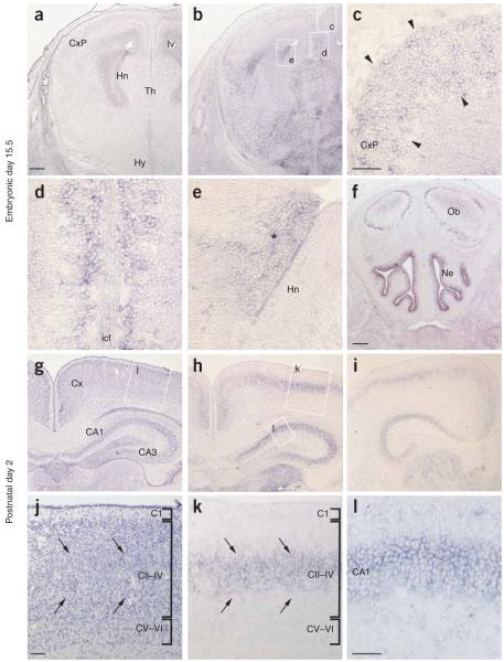
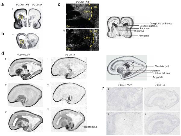
Epilepsy and mental retardation limited to females (EFMR) is a disorder with an X-linked mode of inheritance and an unusual expression pattern. Disorders arising from mutations on the X chromosome are typically characterized by affected males and unaffected carrier females. In contrast, EFMR spares transmitting males and affects only carrier females. Aided by systematic resequencing of 737 X chromosome genes, we identified different protocadherin 19 (PCDH19) gene mutations in seven families with EFMR. Five mutations resulted in the introduction of a premature termination codon. Study of two of these demonstrated nonsense-mediated decay of PCDH19 mRNA. The two missense mutations were predicted to affect adhesiveness of PCDH19 through impaired calcium binding. PCDH19 is expressed in developing brains of human and mouse and is the first member of the cadherin superfamily to be directly implicated in epilepsy or mental retardation.


Figures

Figure 1
Figure 1
Mutations in PCDH19 cause EFMR. Pedigrees of the seven EFMR families showing the characteristic inheritance pattern of affected females and transmitting males. Each of the X chromosome-encoded PCDH19 mutations segregated with the EFMR clinical phenotype. An example of a sequence chromatogram of a PCDH19 mutation as detected in an affected female is shown for each family.
Figure 2
Figure 2
Structure and expression analysis of PCDH19. (a) Schematic diagram of the PCDH19 protein showing the signal peptide, extracellular cadherin (EC), transmembrane (TM) and cytoplasmic (CM) domains. The positions of the mutations found in the families bearing EFMR are shown. (b) Partial alignment of human PCDHs and orthologs of PCDH19 from other species, showing the high conservation of the residues affected by the two missense mutations, V441E (top) and N557K (bottom). Asn557 is invariant and one of the essential residues for calcium ion binding,. Val441 is highly conserved and in close proximity to the calcium-binding acidic residues (bracket). (c) RNA blot analyses of PCDH19 and PCDH11X/Y in various human tissues. Asterisks, the ~9.8 kb PCDH19 transcripts; arrowheads, the smaller, ~9.5 kb PCDH11X/Y mRNAs. Brackets, either nonspecific binding of the PCDH19 probe or PCDH19 degradation products, as there is no evidence for such alternative isoforms of PCDH19 available from our data (data not shown) or from published data. (d) Nonsense-mediated RNA decay of mutant PCDH19 transcripts. Sequence chromatogram from EFMR-affected female from family 2 showing the detection of the mutation 253C>T in genomic DNA (gDNA) (top), the absence of the mutant sequence in fibroblast cDNA (middle) and the presence of the both mutant and wild-type cDNA after the treatment of fibroblasts with cycloheximide (bottom), which inhibits the pioneer round of translation and thus NMD. The position of the mutation is boxed.
Figure 3
Figure 3
Expression of Pcdh19 in the developing mouse CNS. (a-l) Expression at embryonic day 15.5 (a-f) and postnatal day 2 (g-l) (representative of two males and two females studied). (a,b) Adjacent coronal sections through the hippocampal region stained with hematoxylin and eosin or processed for Pcdh19 in situ, respectively. (c-e) Higher magnification images of the boxed regions in b. Arrowheads in c, Pcdh19-expressing cells within the cortex; asterisk in e, dorsolateral wall of the lateral ventricle. (f) Coronal section through the olfactory bulb highlighting Pchd19 expression in the nasal epithelium. (g,h) Adjacent coronal sections through the mid-hippocampal region stained with hematoxylin and eosin or processed for Pcdh19 in situ, respectively. (i) A brain section more posterior than that in h, highlighting Pcdh19 expression. (j-l) Higher-magnification images of the regions boxed in g and h, as indicated. Arrows in j,k, Pcdh19 expression within cortical layers II-IV. Cx, cortex; CxP, cortical plate; Hn, hippocampal neuroepithelium; lv, lateral ventricle; Th, thalamus; Hy, hypothalamus; icf, intercerebral fissure; Ob, olfactory bulbs; Ne, nasal epithelium. Scale bars in a,b,f-i, 200 μM; in c-e,j-l, 50 μM.
Figure 4
Figure 4
Expression of PCDH11X/Y and PCDH19 in midgestation developing human CNS. (a,b) Autoradiography in situ hybridization of female (f; panel a) and male (m; panel b) coronal sections through the basal regions of the brain using probes for PCDH11X/Y and PCDH19, showing several areas of overlap and differences, including high expression of PCDH11X/Y in the ganglionic eminence and sexually dimorphic expression in the caudate nucleus. Dark color denotes high expression. Both genes are highly expressed in amygdala and developing cortical plate. A cartoon key to these sections is shown at the far right and the boxed regions are shown magnified in c. (c) Micrographs from photographic emulsions of magnified regions of PCDH11X/Y expression show low expression in male caudate. White denotes strong expression. GE, ganglionic eminence; Put, putamen; CaNu, caudate nucleus. (d) Sagittal photomicrographs highlight areas of differential expression between the two PCDH genes, indicating high putamen expression for PCDH11X/Y and high hippocampal expression for PCDH19. We show sections from two different planes in the male, the first more lateral and the second more medial, to show that the absence of male expression is not an artifact of section plane. (e) Sections hybridized with sense probes show absence of specific staining.

Comment in

References

    1. Ryan SG, et al. Epilepsy and mental retardation limited to females: an X-linked dominant disorder with male sparing. Nat. Genet. 1997;17:92–95. - PubMed
    1. Juberg RC, Hellman CD. A new familial form of convulsive disorder and mental retardation limited to females. J. Pediatr. 1971;79:726–732. - PubMed
    1. Fabisiak K, Erickson RP. A familial form of convulsive disorder with or without mental retardation limited to females: extension of a pedigree limits possible genetic mechanisms. Clin. Genet. 1990;38:353–358. - PubMed
    1. Scheffer IE, et al. Epilepsy and mental retardation limited to females: an under-recognised disorder. Brain. 2008;131:918–927. - PubMed
    1. Wolverton T, Lalande M. Identification and characterization of three members of a novel subclass of protocadherins. Genomics. 2001;76:66–72. - PubMed

Publication types

MeSH terms