. 2008 Aug 28;27(37):5045-56.
doi: 10.1038/onc.2008.139. Epub 2008 May 12.

Snm1B/Apollo mediates replication fork collapse and S Phase checkpoint activation in response to DNA interstrand cross-links

Affiliations

Snm1B/Apollo mediates replication fork collapse and S Phase checkpoint activation in response to DNA interstrand cross-links

J-B Bae et al. Oncogene. .

Abstract

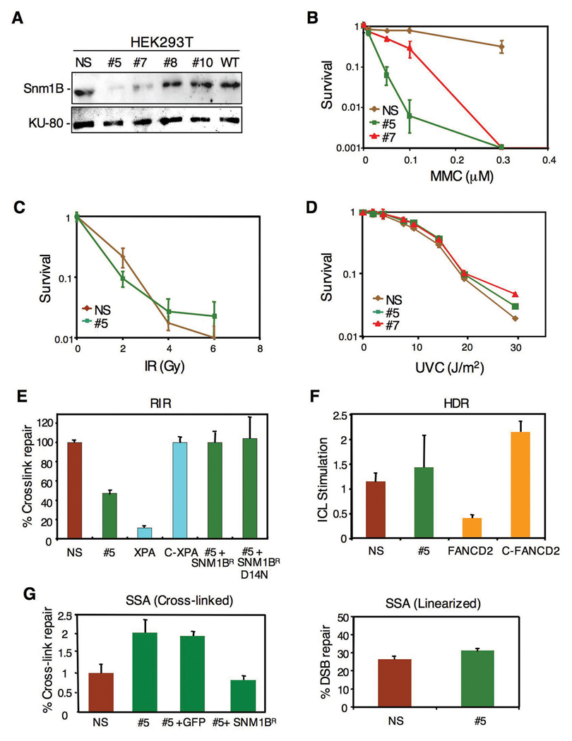
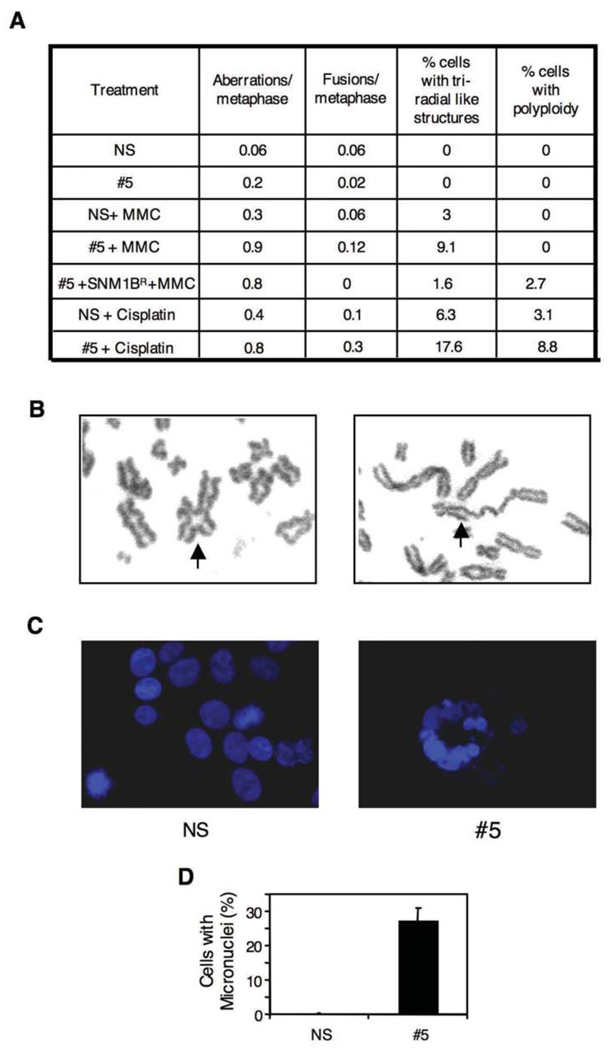
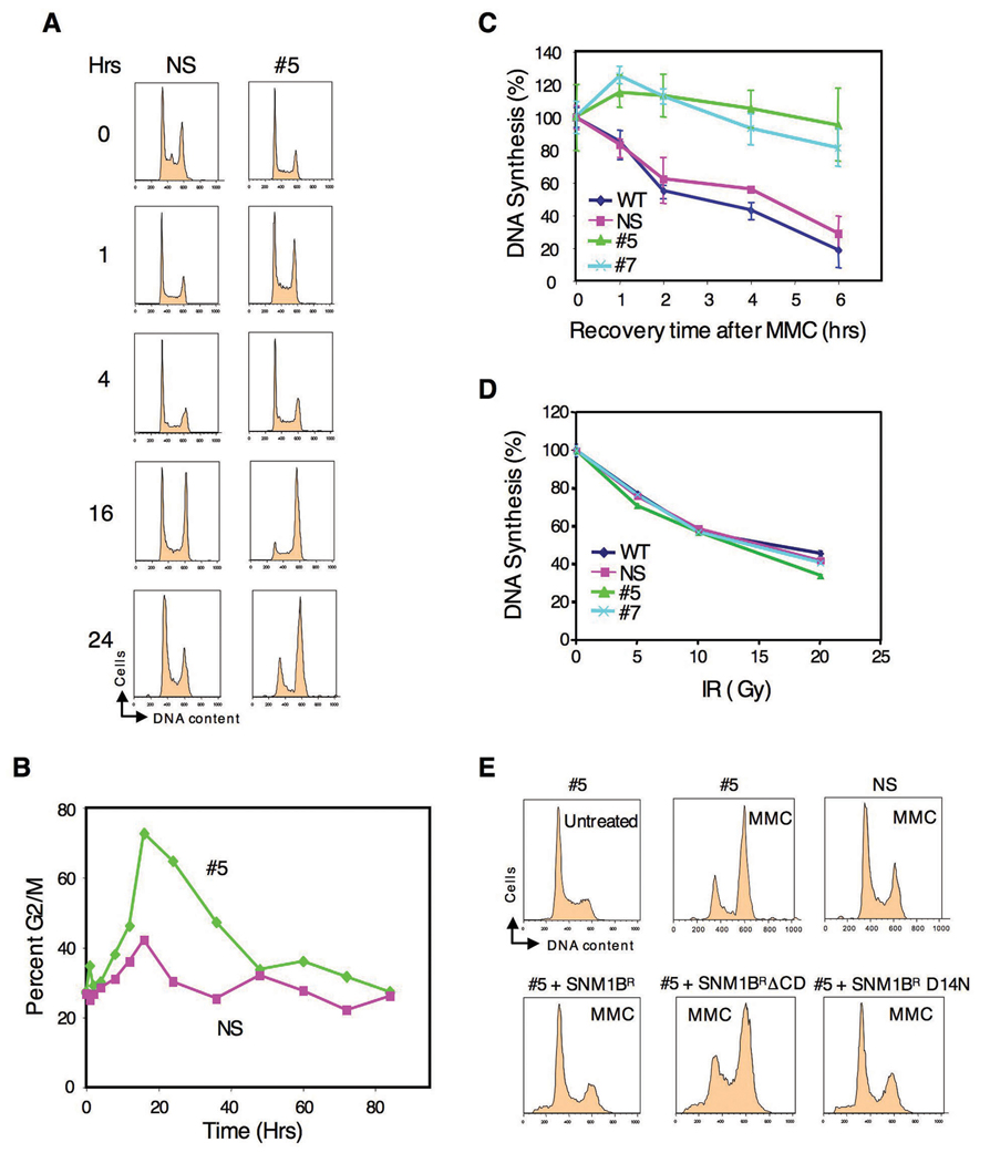
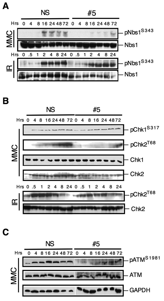
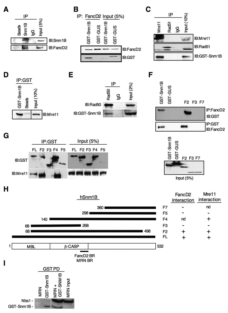
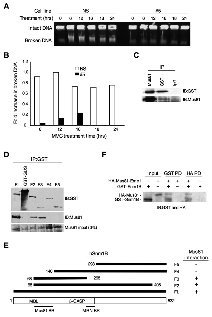
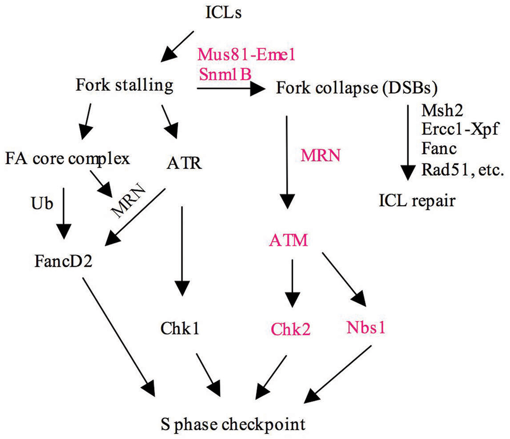
The removal of DNA interstrand cross-links (ICLs) has proven to be notoriously complicated due to the involvement of multiple pathways of DNA repair, which include the Fanconi anemia/BRCA pathway, homologous recombination and components of the nucleotide excision and mismatch repair pathways. Members of the SNM1 gene family have also been shown to have a role in mediating cellular resistance to ICLs, although their precise function has remained elusive. Here, we show that knockdown of Snm1B/Apollo in human cells results in hypersensitivity to mitomycin C (MMC), but not to IR. We also show that Snm1B-deficient cells exhibit a defective S phase checkpoint in response to MMC, but not to IR, and this finding may account for the specific sensitivity to the cross-linking drug. Interestingly, although previous studies have largely implicated ATR as the major kinase activated in response to ICLs, we show that it is activation of the ATM-mediated checkpoint that is defective in Snm1B-deficient cells. The requirement for Snm1B in ATM checkpoint activation specifically after ICL damage is correlated with its role in promoting double-strand break formation, and thus replication fork collapse. Consistent with this result Snm1B was found to interact directly with Mus81-Eme1, an endonuclease previously implicated in fork collapse. In addition, we also show that Snm1B interacts with the Mre11-Rad50-Nbs1 (MRN) complex and with FancD2 further substantiating its role as a checkpoint/DNA repair protein.


Figures

Figure 1
Figure 1. Knockdown of Snm1B by RNAi causes sensitivity to DNA interstrand cross-linking agents, but Snm1B is not involved in the recombination steps of ICL repair
(A) Stable knockdown of Snm1B by shRNA in HEK293T cells as shown by immunoblotting. Numbered clones with various levels of knockdown are shown. WT indicates untransfected wild-type cells. NS indicates a clone expressing a nonspecific shRNA. (B–D) Colony survival assays for clones #5, #7, and NS following exposure to the indicated DNA damaging treatments. (E) SNM1B-deficient cells are partially defective in recombination-independent DNA repair (RIR). Reactivation of a luciferase reporter gene by repair of a single site-specific psoralen ICL in the indicated cell lines is shown. The relative efficiencies were calculated as the percentage of luciferase activity of the cross-linked reporter gene normalized to that of unmodified reporter gene. C-XPA indicates a clone with stable correction of the XPA cell line. SNM1BR indicates an allele refractory to the SNM1B shRNA. D14N indicates a point mutation of SNM1B in the metallo-β-lactamase domain. All assays were carried out in triplicate and standard deviations are indicated. (F) SNM1B-deficient cells show no defect in homology dependent recombination (HDR) of a linearized cross-linked substrate. pECFPHR contains a donor ECFP gene without a start codon and an interrupted ECFP gene due to the insertion of an oligonucleotide, with or without a psoralen cross-link, within the coding region (Zhang et al., 2007). The ratio of homologous recombination in cross-linked plasmids to homologous recombination in noncross-linked plasmids is presented as ICL stimulation of HDR. (G) SNM1B-deficient cells exhibit increased levels of repair by SSA. Psoralen-crosslinked (left panel) or non-crosslinked, linearized (right panel) pSupN plasmids were transfected into the indicated cell lines. For the rescue experiment the control (GFP) and SNM1BR DNAs were co-transfected with the pSupN plasmid. Recombination frequency refers to percentage of blue colonies derived from more than 10,000 total colonies.
Figure 2
Figure 2. Knockdown of Snm1B causes chromosomal abnormalities
(A) Quantification of various chromosomal aberrations after MMC (50 ng/ml) or cisplatin (2 µM) treatment. Cells were treated with the indicated agent for 8 hrs and then allowed to recover for 24 hrs in fresh medium. Colcemid was then added for 1 hr to accumulate mitotic cells. (B) Examples of aberrant chromosomes scored as indicated in (A). Arrows indicate a triradial (left panel) and a fused chromosome (right panel). (C) Knockdown of Snm1B induces micronuclei formation after treatment with MMC (50 ng/ml). Nuclei are stained with DAPI (blue). (D) Quantification of micronuclei shown in (C).
Figure 3
Figure 3. Cell cycle analysis of SNM1B knockdown cells after MMC treatment
(A) Knockdown of Snm1B results in G2/M accumulation in the presence of MMC. The indicated clones were continuously exposed to MMC (100 ng/ml) and examined at the indicated time points for DNA content by FACS analysis. (B) Quantification of the G2/M accumulation shown in (A). (C,D) SNM1B-deficient cells are defective in an S phase checkpoint after treatment with MMC, but not with IR. DNA synthesis was evaluated (as described in Experimental Procedures) with the indicated clones after treatment with MMC (10 µg/ml for 1 hr), or as a function of IR dose. (E) The hydrolase activity of Snm1B is not required for rescue of the G2/M accumulation phenotype. The indicated clones were either treated with MMC (50 ng/ml) for 24 hrs or not treated, and then analyzed by FACS. For the rescue experiments (lower panels), the indicated SNM1B constructs were transfected into #5 cells, and 24 hrs later MMC was added for an additional 24 hrs. The Snm1B proteins were tagged with EGFP and only the GPF positive cell populations as determined by FACS analysis are shown. The SNM1B alleles are as described in Fig. 3A, except for SNM1ΔCD which lacks the entire conserved domain (metallo-β-lactamase plus β-CASP).
Figure 4
Figure 4. SNM1B is required for checkpoint signaling through the ATM pathway
(A) Phosphorylation of Nbs1 is defective in SNM1B knockdown cells upon exposure to MMC. The NS and #5 clones were treated with MMC (50 ng/ml) continuously or to IR (2 Gy), and harvested at the indicated time points for immunoblotting. (B) Phosphorylation of Chk2, but not Chk1, is defective in SNM1B knockdown cells upon exposure to MMC. The NS and #5 clones were continuously exposed to MMC (300 ng/ml) or to IR (2 Gy) for the indicated times. (C) Activation of ATM is defective in SNM1B knockdown cells. The NS and #5 clones were continuously exposed to MMC (300 ng/ml) for the indicated times.
Figure 5
Figure 5. Snm1B interacts with FancD2 and the MRN complex
(A) Antibodies to Snm1B co-IP FancD2. GST-Snm1B was transiently expressed in HEK293 cells, and the indicated co-IP assays were performed from lysates. “Beads” indicates IP with sepharose A beads only. “IgG” indicates IP with nonspecific antibody. (B) Antibodies to FancD2 co-IP Snm1B. GST-Snm1B or the control GST-GUS were transiently expressed in HEK293 cells, and co-IP assays were performed from lysates. (C) Antibodies to Mre11, but not Rad51, co-IP Snm1B. GST-Snm1B was transiently expressed in HEK293 cells, and the indicated co-IP assays were performed from lysates. “IgG” indicates IP with nonspecific antibody. (D) Reciprocal co-IP of Mre11 with antibodies to GST. “Beads” indicates IP with sepharose A beads only. (E) Snm1B co-IPs with Rad50. GST-Snm1B was transiently expressed in HEK293 cells, and the indicated co-IP assays were performed from lysates. (F) The FancD2 interaction domain maps to the carboxy terminal end of the β-CASP domain of Snm1B. The indicated GST-SNM1B deletion constructs were transiently expressed in HEK293 cells, and the indicated co-IP assays were performed. “FL” indicates full-length Snm1B. (G) The Mre11 interaction domain maps to the carboxy terminal end of the β-CASP domain of Snm1B. The indicated GST-SNM1B deletion constructs were transiently expressed in HEK293 cells and the indicated co-IP assays were performed. (H) Schematic depicting the interaction mapping results from (F) and (G). “nd” indicates not determined. “BR” indicates binding region. (I) Snm1B interacts directly with the MRN complex. Purified recombinant GST-Snm1B and MRN complex were incubated together, and then subjected to the indicated pull-down (PD) assay. Immunoblotting was performed with anti-GST and anti-Nbs1.
Figure 6
Figure 6. Snm1B is required for fork collapse after MMC treatment
(A) Double-strand break formation was analyzed by pulsed field gel electrophoresis. Intact DNA stays in the well while broken DNA migrates into the agarose gel. The NS and #5 clones were incubated with MMC (1 ug/ml) for the indicated times, and cells were collected in agarose plugs for gel electrophoresis. (B) Quantitation of results shown in (A). Images were quantitated using ImageJ 1.37v software (developed by Wayne Rasband, NIH). The background present at the 0 hr time point was subtracted from the other time points, and the band with the highest intensity was normalized to a value of 1.0. (C) Snm1B co-IPs with Mus81. GST-Snm1B was transiently expressed in HEK293 cells, and the indicated co-IP assays were performed from lysates. (D) The Mus81 interaction domain maps to the metallo-β-lactamase domain of Snm1B. The indicated GST-SNM1B deletion constructs were expressed in HEK293 cells and the indicated co-IP assays were performed and analyzed by immunoblotting. (E) Schematic depicting the results from (D). (F) Snm1B directly interacts with Mus81-Eme1. Immunoblots showing results of reciprocal pull-down (PD) assays between GST-Snm1B and HA-Mus81-Eme1. HA indicates the hemagglutinin tag.
Figure 7
Figure 7. Model depicting the role of Snm1B in the cellular response to ICLs
Items in bold indicate pathways likely affected by Snm1B.

References

    1. Ahkter S, Richie CT, Zhang N, Behringer RR, Zhu C, Legerski RJ. Snm1-deficient mice exhibit accelerated tumorigenesis and susceptibility to infection. Mol Cell Biol. 2005;25:10071–10078. - PMC - PubMed
    1. Akhter S, Richie CT, Deng JM, Brey E, Zhang X, Patrick, et al. Deficiency in SNM1 abolishes an early mitotic checkpoint induced by spindle stress. Mol Cell Biol. 2004;24:10448–10455. - PMC - PubMed
    1. Akkari YM, Bateman RL, Reifsteck CA, D'Andrea AD, Olson SB, Grompe M. The 4N cell cycle delay in Fanconi anemia reflects growth arrest in late S phase. Mol Genet Metab. 2001;74:403–412. - PubMed
    1. Andreassen PR, D'Andrea AD, Taniguchi T. ATR couples FANCD2 monoubiquitination to the DNA-damage response. Genes Dev. 2004;18:1958–1963. - PMC - PubMed
    1. Bakkenist CJ, Kastan MB. DNA damage activates ATM through intermolecular autophosphorylation and dimer dissociation. Nature. 2003;421:499–506. - PubMed

Publication types

MeSH terms