. 1991 Jan 14;278(1):107-14.
doi: 10.1016/0014-5793(91)80095-k.

Complete cDNA sequences of mouse rod photoreceptor cGMP phosphodiesterase alpha- and beta-subunits, and identification of beta'-, a putative beta-subunit isozyme produced by alternative splicing of the beta-subunit gene

Affiliations

Complete cDNA sequences of mouse rod photoreceptor cGMP phosphodiesterase alpha- and beta-subunits, and identification of beta'-, a putative beta-subunit isozyme produced by alternative splicing of the beta-subunit gene

W Baehr et al. FEBS Lett. .

Abstract

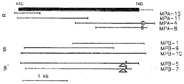
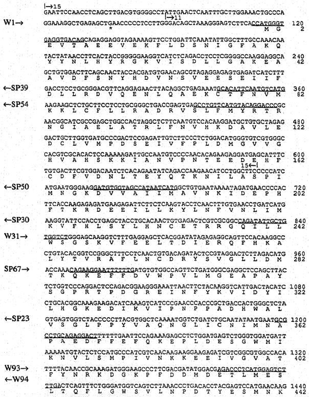
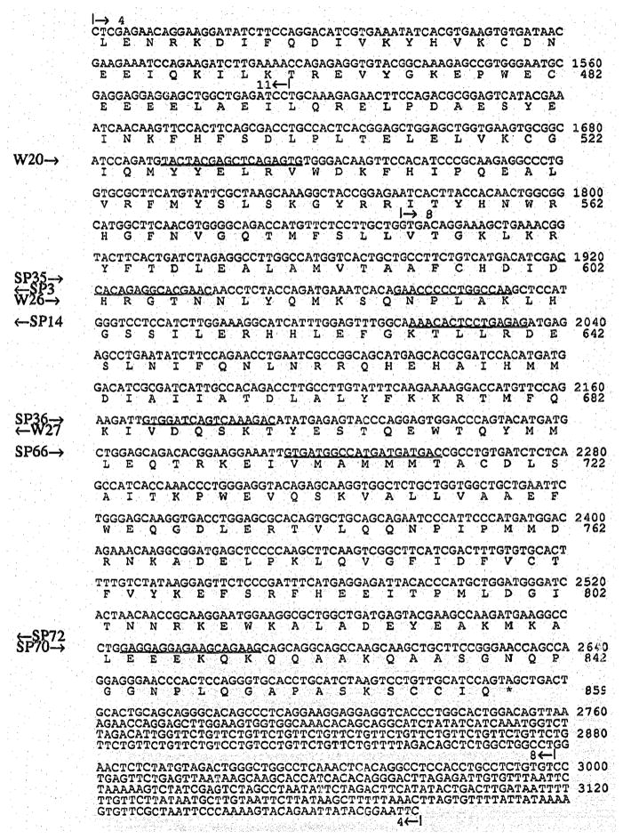
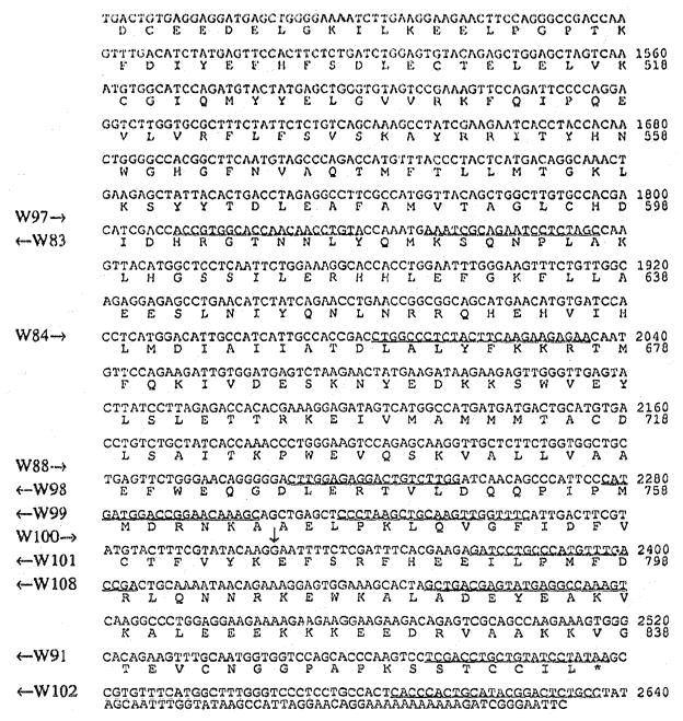
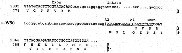
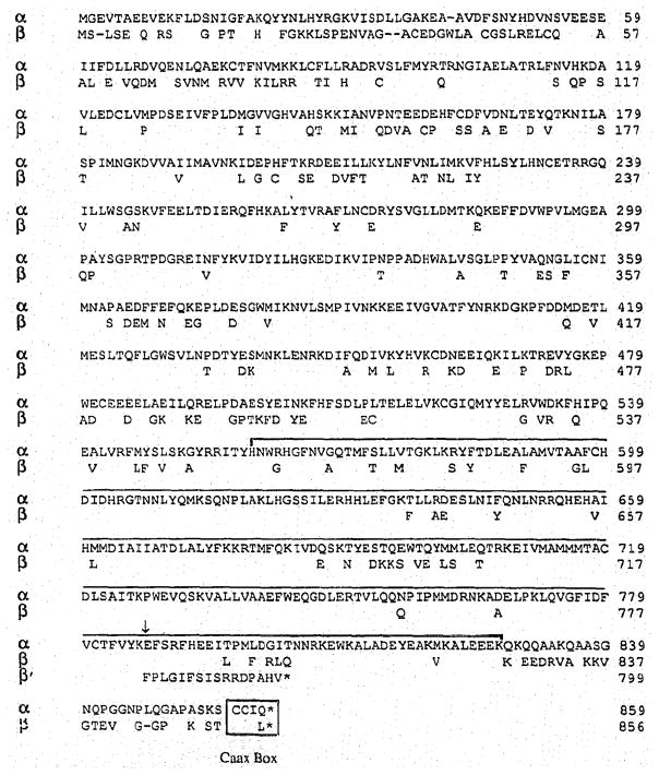
We have characterized overlapping cDNA clones encoding cGMP phosphodiesterase (PDE) alpha- and beta-subunits of mouse retinal rod photoreceptors. The open reading frames predict an alpha-subunit of 100 kDa (856 residues), and a beta-subunit of 99 kDa (853 residues). Sequence analysis of two of twelve beta-subunit clones predicts the presence in the retina of an additional PDE, termed beta', which is generated by alternative splicing of the beta-subunit gene. beta' differs from beta only at the C-terminus being 55 residues shorter and lacking the Caax motif found at the C-termini of both the alpha- and beta-subunits. A 300 residue segment thought to contain the active site is present in the C-terminal half of alpha, beta and beta'.


Figures

Fig. 1
Fig. 1
Map of cDNA clones encoding the mouse PDE α-, β-and β′-polypeptides. The black box depicts the coding sequences of the α- and β-subunit. The extent of clones is indicated by bold-faced lines. The rectangles in the 3′ untranslated region of MPA-4 and MPA-8 represent (TTCTG)n repeats, where n = 17 in MPA-4 and n = 9 in MPA-8. The triangles in MPB-5 and MPB-7 symbolize a 10 bp insertion. The asterisk indicates the location of the first in-frame stop codon.
Fig. 2
Fig. 2
cDNA and predicted amino acid sequence of the mouse PDE α-subunit. Primers used for DNA sequencing are underlined, and their sense or antisense direction is indicated on the left margin by arrows pointing to the right or left. Start and end points of overlapping clones (Fig. 1) are shown above the sequence.
Fig. 2
Fig. 2
cDNA and predicted amino acid sequence of the mouse PDE α-subunit. Primers used for DNA sequencing are underlined, and their sense or antisense direction is indicated on the left margin by arrows pointing to the right or left. Start and end points of overlapping clones (Fig. 1) are shown above the sequence.
Figure 3
Figure 3
Fig. 3A. cDNA and predicted amino acid sequences of the mouse PDE β-subunit. The start point of clone MPB-10 is indicated above the sequence. Primers used for DNA and RNA sequencing, and for PCR amplification are underlined. The vertical arrow indicates the 10 bp insertion point. Fig. 3B. Junctions of the 1.4 kb intron that is alternatively spliced. Predicted amino acid sequences of the β-subunit and the β′-isozyme are shown below the sequence. The numbering on the left is according to Fig. 3A. W90 is an antisense, insertion specific primer. A1, junction one at the 3′ end of the intron used for generation of β; A2, junction two used for β′.
Figure 3
Figure 3
Fig. 3A. cDNA and predicted amino acid sequences of the mouse PDE β-subunit. The start point of clone MPB-10 is indicated above the sequence. Primers used for DNA and RNA sequencing, and for PCR amplification are underlined. The vertical arrow indicates the 10 bp insertion point. Fig. 3B. Junctions of the 1.4 kb intron that is alternatively spliced. Predicted amino acid sequences of the β-subunit and the β′-isozyme are shown below the sequence. The numbering on the left is according to Fig. 3A. W90 is an antisense, insertion specific primer. A1, junction one at the 3′ end of the intron used for generation of β; A2, junction two used for β′.
Fig. 4
Fig. 4
Alignment of mouse PDE α- and β-subunit amino acid sequences. Gaps are indicated by a hyphen, identical residues by blanks. The C-terminal 16 residues of β′ are shown from the point (arrow) where the frame shift occurs in the cDNA sequence. An asterisk depicts a stop codon. The conserved domain thought to be involved in catalysis is bracketed. The Caax motif at the C-terminus is boxed.

References

    1. Al-Ubaidi MR, Pittler SJ, Champagne MS, Triantafyllos JT, McGinnis JF, Baehr W. J Biol Chem. 1990:265. in press. - PubMed
    1. Badley JE, Bishop GA, St John T, Frelinger JA. BioFeedback. 1988;6:114–116. - PubMed
    1. Baehr W, Devlin MJ, Applebury ML. J Biol Chem. 1979;254:11669–11677. - PubMed
    1. Beavo JA. Adv Second Messenger Phosphoprotein Res. 1988;22:1–38. - PubMed
    1. Bowes C, Danciger M, Kozak CA, Farber DB. Proc Natl Acad Sci USA. 1989;86:9722–9726. - PMC - PubMed

Publication types