. 2008 Jun 15;318(2):323-34.
doi: 10.1016/j.ydbio.2008.03.037. Epub 2008 Apr 8.

Twist1 homodimers enhance FGF responsiveness of the cranial sutures and promote suture closure

Affiliations

Twist1 homodimers enhance FGF responsiveness of the cranial sutures and promote suture closure

Jeannette Connerney et al. Dev Biol. .

Erratum in

  • Dev Biol. 2012 Jun 15;366(2):434

Abstract

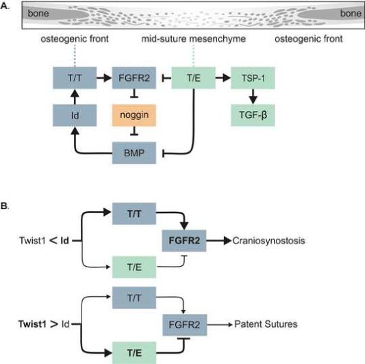
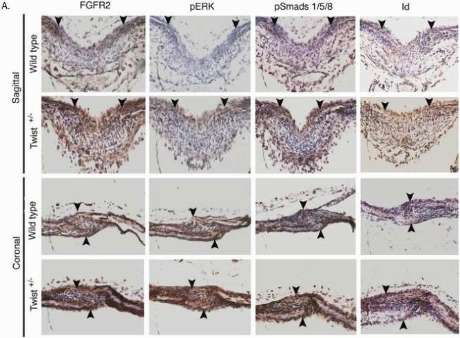
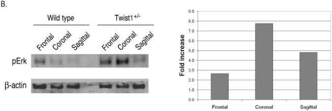
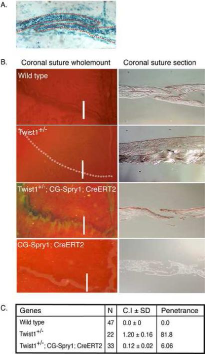
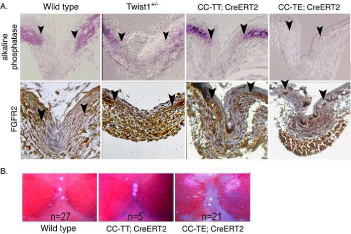
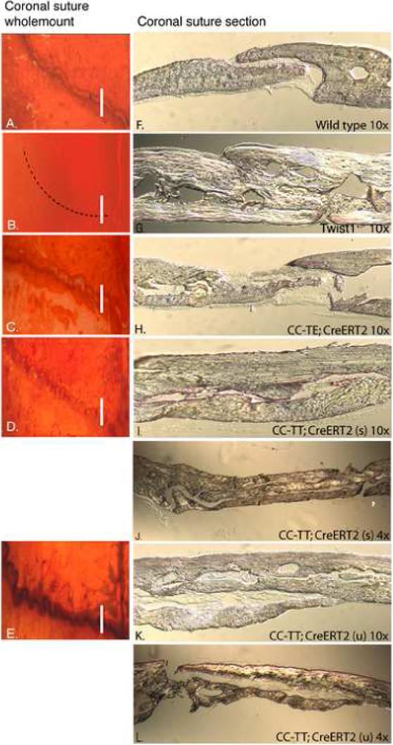
Haploinsufficiency of the transcription factor TWIST1 is associated with Saethre-Chotzen Syndrome and is manifested by craniosynostosis, which is the premature closure of the calvaria sutures. Previously, we found that Twist1 forms functional homodimers and heterodimers that have opposing activities. Our data supported a model that within the calvaria sutures Twist1 homodimers (T/T) reside in the osteogenic fronts while Twist1/E protein heterodimers (T/E) are in the mid-sutures. Twist1 haploinsufficiency alters the balance between these dimers, favoring an increase in homodimer formation throughout the sutures. The data we present here further supports this model and extends it to integrate the Twist1 dimers with the pathways that are known to regulate cranial suture patency. This data provides the first evidence of a functional link between Twist1 and the FGF pathway, and indicates that differential regulation of FGF signaling by T/T and T/E dimers plays a central role in governing cranial suture patency. Furthermore, we show that inhibition of FGF signaling prevents craniosynostosis in Twist1(+/-) mice, demonstrating that inhibition of a signaling pathway that is not part of the initiating mutation can prevent suture fusion in a relevant genetic model of craniosynostosis.


Figures

Figure 1
Figure 1
Model. (A) Drawing showing parts of a suture going from a more differentiated bone to a less differentiated mid-suture mesenchyme. The model shows T/T dimers up-regulating FGFR2 expression resulting in increased FGF signaling toward the middle of the suture. FGF signaling inhibits the expression of the BMP antagonist noggin, resulting in increased BMP signaling which up-regulates Id1 expression, further enhancing T/T homodimer formation. On the other hand, T/E dimers inhibit FGFR2 expression, as well as BMP signaling through binding to Smad proteins. T/E dimers also up-regulate TSP1 expression, which activates latent TGFβ. This could have differing outcomes dependent on which TGFβ isoform is present. (B) The ratio of T/T to T/E determines the functional output of Twist1 expression, and this is determined by the relative expression of Twist1 and Id1 proteins. When Id1 levels are greater that Twist1 this balance is shifted toward T/T formation resulting in up-regulation of FGFR2 leading to craniosynostosis. When Twist1 levels are higher than Id1 the balance is toward an increase in T/E formation, inhibiting FGFR2 expression and resulting in patent sutures.
Figure 2
Figure 2
Increased FGF and BMP signaling in the sutures of Twist1+/− mice. (A) Immunohistochemistry for FGFR2, phospho-ERK1/2, phospho-smad1/5/8, and Id1 proteins in the sagittal and coronal sutures of wild type and Twist1+/− P1 mice. Arrow heads indicate the osteogenic fronts. Analysis of at least 3 mice of each genotype was performed, and representative data is shown. Note the extended staining for all of these throughout the sutures of Twist1+/− mice, while they are primarily restricted to the osteogenic fronts in wild type mice. (B) Western blot analysis of phopho-Erk1/2 in wild type and Twist1+/− sutures. Protein extracts from dissected frontal, coronal, and sagittal sutures from two P1 wild type or Twist1+/− mice were pooled and analyzed by western blot analysis for phopho-Erk1/2 and β-actin. The bar graph shows the fold change in phospho-Erk1/2 for each suture between wild type and Twist1+/− mice when corrected for β-actin expression. (C) in situ hybridization analysis of erm and noggin expression in the sagittal and coronal sutures of wild type and Twist1+/− P1 mice. The red dotted lines outline the calvaria bones.
Figure 2
Figure 2
Increased FGF and BMP signaling in the sutures of Twist1+/− mice. (A) Immunohistochemistry for FGFR2, phospho-ERK1/2, phospho-smad1/5/8, and Id1 proteins in the sagittal and coronal sutures of wild type and Twist1+/− P1 mice. Arrow heads indicate the osteogenic fronts. Analysis of at least 3 mice of each genotype was performed, and representative data is shown. Note the extended staining for all of these throughout the sutures of Twist1+/− mice, while they are primarily restricted to the osteogenic fronts in wild type mice. (B) Western blot analysis of phopho-Erk1/2 in wild type and Twist1+/− sutures. Protein extracts from dissected frontal, coronal, and sagittal sutures from two P1 wild type or Twist1+/− mice were pooled and analyzed by western blot analysis for phopho-Erk1/2 and β-actin. The bar graph shows the fold change in phospho-Erk1/2 for each suture between wild type and Twist1+/− mice when corrected for β-actin expression. (C) in situ hybridization analysis of erm and noggin expression in the sagittal and coronal sutures of wild type and Twist1+/− P1 mice. The red dotted lines outline the calvaria bones.
Figure 3
Figure 3
Twist1+/− sutures are more responsive to FGF. (A) E16.5 calvarias stained with Alcian Blue and Alizarin Red. Note that wild type and Twist1+/− calvaria are phenotypically the same at this stage. The drawing in the middle shows the location of beads (red dots) that were placed on the osteogenic fronts (OF) or on the mid-sutures (MS). (B) BSA or FGF2-soaked beads were placed on the osteogenic fronts or mid-sutures of wild type and Twist1+/− E16.5 calvaria, and the explants were cultured for 7 days. FGF2 beads placed on the osteogenic fronts induced suture closure in both wild type and Twist1+/− explants, while only the Twist1+/− sutures closed when FGF2 beads were placed on the mid-sutures. (C) Table showing the results of beads placed either on osteogenic fronts or mid-suture of calvaria explants of wild type versus Twist1+/− mice. Suture closure was defined as when the two parietal bones came in contact.
Figure 4
Figure 4
FGF signaling is required for craniosynostosis in Twist1+/− mice. (A) Efficient tamoxifen-induced cre recombination in the sutures. The sutures of 3-day-old pups from R26R reporter mice crossed with CAG-CreERT mice were injected with tamoxifen to induce expression of β-galactosidase from the R26R locus. The mice were then harvested at P7 and the coronal sutures were analyzed for β-galactosidase expression. Note expression throughout the coronal suture. (B) Expression of Spry1 in the sutures of Twist1+/− mice prevents craniosynostosis. Spry1 expression was induced from the CG-Spry1 transgene in CG-Spry1;CAG-CreERT2 mice on a wild type or Twist1+/− background by injection of tamoxifen into the sutures of P3 pups, and the coronal sutures were analyzed at 5 weeks. The skulls were stained with alcian blue and alizarin red. The left panels are wholemount analyses of the coronal sutures. The right panels are cross sections through the coronal sutures indicated by the white lines in the left panels. Representative examples are shown. (C) Table indicating the penetrance and degree of suture fusion. Skulls were assessed using a scoring system in which a suture was assigned a value from 0 to 3: 0 is completely unfused; 1 is 30% fused; 2 is 60% fused; and 3 is 100% fused. Left and right coronal sutures were scored individually for each skull. For each genotype, the scoring mean SEM was determined and was termed the craniosynostosis index (CI). Statistical analysis was performed by one-way analysis of variance. Cre expression alone did not affect the Twist1+/− phenotype, and therefore all of the mice that were Twist1+/− but did not have both the CAG-CreERT2 and CG-Spry1 transgenes, and therefore did not express Spry1, were designated as being Twist1+/−. For the same reason, all mice that were wild type for Twist1 were all considered wild type in computing the CI. The difference between Twist1+/− and Twist1+/−;CG-Spry1;CAG-CreERT2 mice, with activated Spry1 expression, was highly significant (p =0.001). Penetrance was calculated as the percentage of animals that exhibited any coronal suture fusion.
Figure 5
Figure 5
Expression of TT leads to a similar increase in FGFR2 expression and responsiveness as Twist1+/− mice. (A) Twist1 homodimers (CC-TT) promote FGFR2 expression and osteoblast differentiation, while Twist1 heterodimers (CC-TE) inhibit these. All pups were injected with tamoxifen at P3 to induce transgene expression, and the sutures were analyzed at P7. Sections through the sagittal sutures of P7 skulls from wild type, Twist1+/−, CC-TT;CAG-CreERT2, and CC-TE;CAG-CreERT2 mice were analyzed for alkaline phosphatase and FGFR2 protein expression. Arrowheads indicate the osteogenic fronts. Analysis of at least 3 mice of each genotype was performed, and representative data is shown. Note that FGFR2 expression was limited to the osteogenic fronts in wild type sutures, while Twist1+/− and CC-TT;CAG-CreERT2 sutures had an expansion of this expression to the middle of the suture. Mice expressing CC-TE had even lower FGFR2 expression than wild type. Alkaline phosphatase staining was slightly expanded from the osteogenic fronts in Twist1+/− sutures, but was significantly enhanced in CC-TT;CAG-CreERT2 mice and almost absent in CC-TE;CAG-CreERT2 mice. (B) FGF responsiveness is enhanced by TT and decreased by TE expression. Pregnant females were injected with tamoxifen at E15 and then at E16.5 calvaria explants of wild type, CC-TT;CAG-CreERT2, and CC-TE;CAG-CreERT2 were incubated for 5 days in differentiation media with FGF2 soaked beads placed on the mid-suture. FGF beads placed on the mid-suture of CC-TT-expressing calvaria promoted suture closure in all of the explanted calvaria, which did not occur in any of the control or CC-TE-expressing explants.
Figure 6
Figure 6
Expression of TT promotes overgrowth of the calvaria bones. (A-L) Mice were treated at P3 as in Figure 4 and were then harvested for analysis at 5 weeks of age. Skulls were stained with alcian blue and alizarin red and analyzed by wholemount (A-E) and sectioning (F-L). The number of mice treated was wild type (n=9), CC-TE (n=19), and CC-TT (n=8). Coronal sutures appeared patent in wild type mice (A), while Twist1+/− sutures were fused (B). TT expression resulted in varied degrees of two phenotypes: CC-TT (u) presented with an undulate phenotype (D), and CC-TT (s) had a serrate phenotype, which appeared to be tightly closed (E). The TE-expressing sutures were patent and showed greater separation between the bones (C). Cross-sections of coronal sutures confirmed the difference between the sutures: (F) coronal sutures from wild type adult mice showed a defined opening between the parietal and frontal bones; (G) Twist1+/− coronal sutures were clearly fused and the resulting bone was thicker than wild type; (H) CC-TE;CAG-CreERT2 sutures showed an increased mid-suture space with thinner bone ends compared to wild type; (I-L) CC-TT;CAG-CreERT2 expressing sutures of both serrate (s) (I,J) and undulate (u) (K,L) phenotypes were significantly expanded with severely overgrown calvaria bones that resembled the thickness of the Twist1+/− calvaria. These sutures are also shown using 4× magnification objectives to be able to view the entire sutures (J,L).

References

    1. Annes JP, et al. Making sense of latent TGFbeta activation. J Cell Sci. 2003;116:217–24. - PubMed
    1. Araki K, et al. Site-specific recombination of a transgene in fertilized eggs by transient expression of Cre recombinase. Proc Natl Acad Sci U S A. 1995;92:160–4. - PMC - PubMed
    1. Bialek P, et al. A twist code determines the onset of osteoblast differentiation. Dev Cell. 2004;6:423–35. - PubMed
    1. Brent AE, Tabin CJ. FGF acts directly on the somitic tendon progenitors through the Ets transcription factors Pea3 and Erm to regulate scleraxis expression. Development. 2004;131:3885–96. - PubMed
    1. Carver EA, et al. Craniosynostosis in Twist heterozygous mice: A model for Saethre-Chotzen syndrome. Anat Rec. 2002;268:90–2. - PubMed

Publication types

Substances

LinkOut - more resources