. 2008 Jun 13;379(4):859-70.
doi: 10.1016/j.jmb.2008.04.013. Epub 2008 Apr 10.

Hinge stiffness is a barrier to RNA folding

Affiliations

Hinge stiffness is a barrier to RNA folding

Jörg C Schlatterer et al. J Mol Biol. .

Abstract

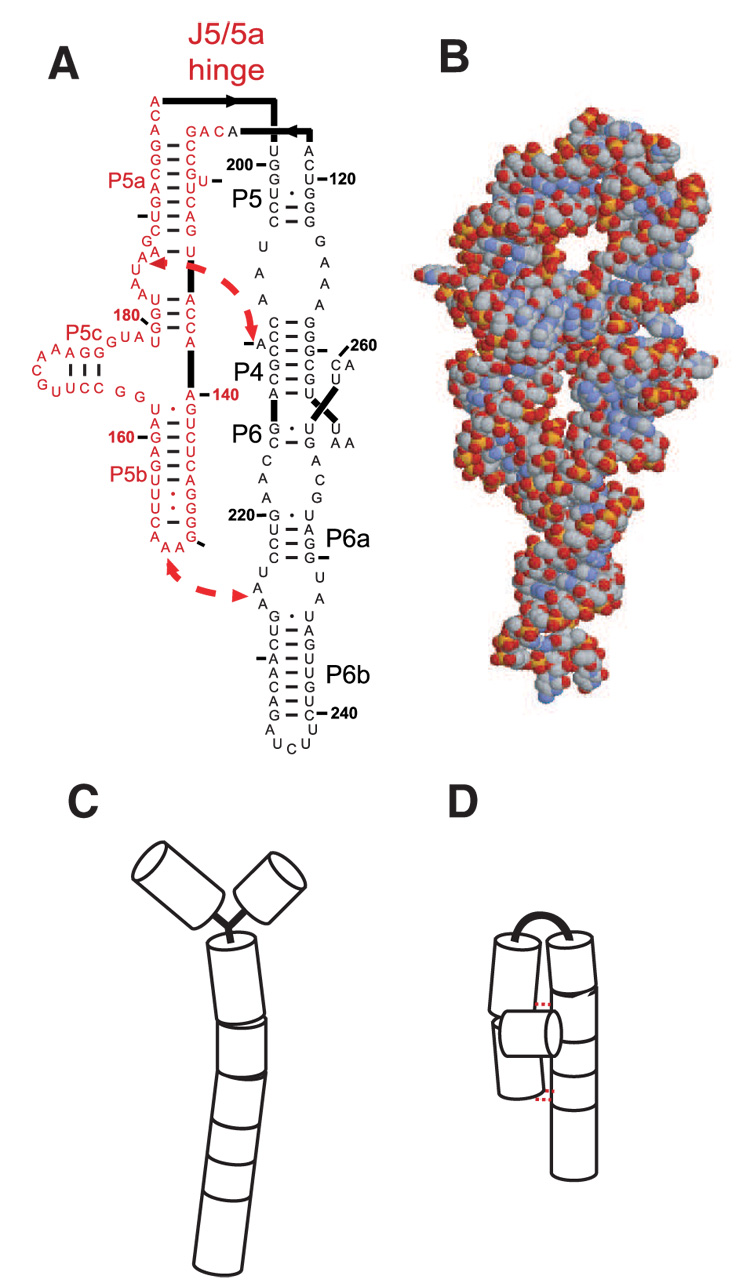
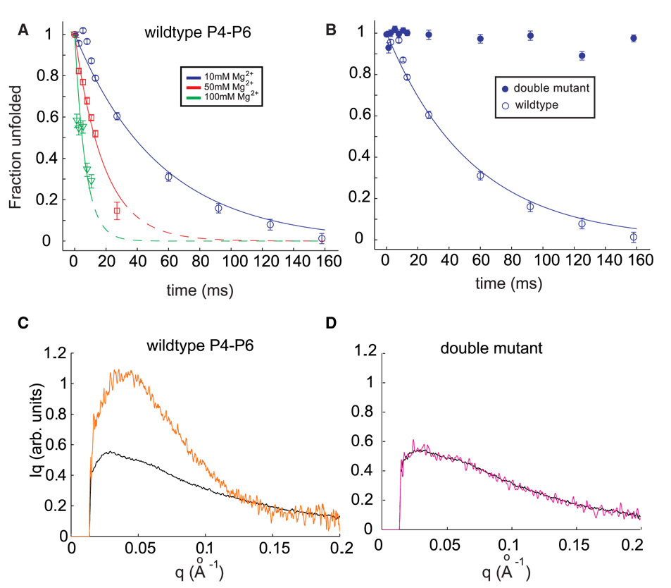
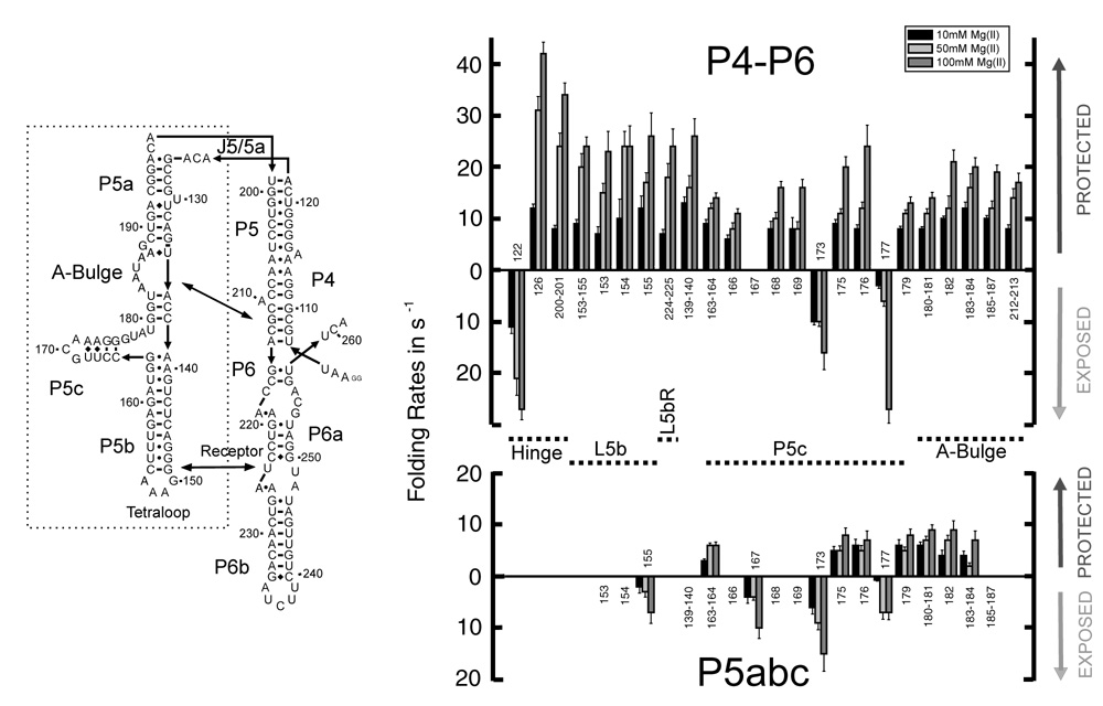
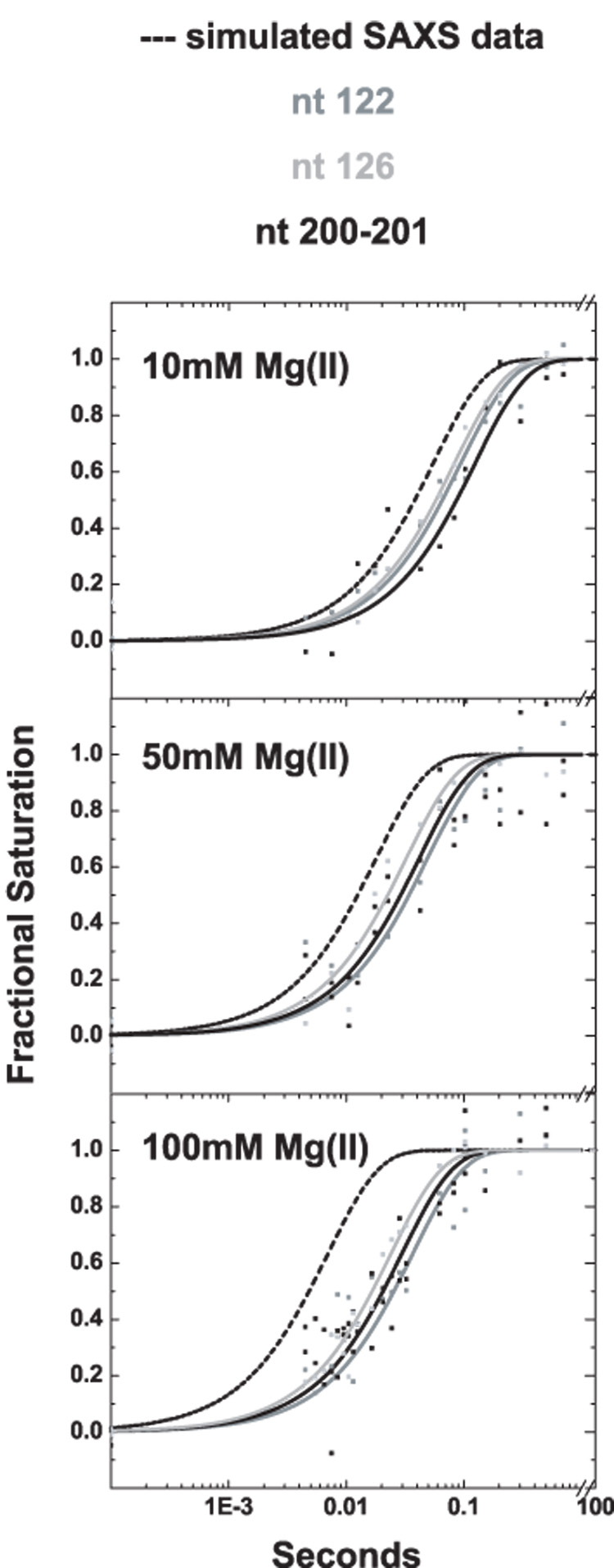
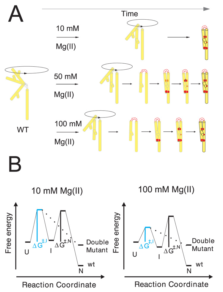
Cation-mediated RNA folding from extended to compact, biologically active conformations relies on a temporal balance of forces. The Mg2 +-mediated folding of the Tetrahymena thermophila ribozyme is characterized by rapid nonspecific collapse followed by tertiary-contact-induced compaction. This article focuses on an autonomously folding portion of the Tetrahymena ribozyme, its P4-P6 domain, in order to probe one facet of the rapid collapse: chain flexibility. The time evolution of P4-P6 folding was followed by global and local measures as a function of Mg2 + concentration. While all concentrations of Mg2 + studied are sufficient to screen the charge on the helices, the rates of compaction and tertiary contact formation diverge as the concentration of Mg2 + increases; collapse is greatly accelerated by Mg2 +, while tertiary contact formation is not. These studies highlight the importance of chain stiffness to RNA folding; at 10 mM Mg2 +, a stiff hinge limits the rate of P4-P6 folding. At higher magnesium concentrations, the rate-limiting step shifts from hinge bending to tertiary contact formati


Figures

Figure 1
Figure 1
The P4–P6 domain. (A) A two dimensional representation showing secondary structure. The locations of the two tertiary contacts are indicated by the large arrows. In the mutant RNA, the tertiary contacts are disrupted by the following sequence modifications: nucleotides AAUAAG (183–188) are replaced by UUUUUU and GGAAAC (149–154) are replaced by CUUCGG. Bases comprising the P5abc sub-domain are shown in red. (B) Reconstruction of the P4–P6 domain based on a monomeric subunit of 1GID.pdb generated by RasMol. (C & D) Simplified models of the extended, unfolded and compact, folded structures, respectively following the representation of Ref. .
Figure 2
Figure 2
(A) Folding time courses for wild type P4–P6 at different magnesium concentrations. The coefficient Pu, representing the percentage of the unfolded scattering profile required to fit each time-resolved profile (see text) is shown following the addition of 10 mM Mg2+ (circles), 50 mM Mg2+ (squares) and 100 mM Mg2+ (triangles). Changes in the time courses can be explained by a model in which the flexibility of the J5-5a hinge varies with ionic strength. (B) The folding time course following addition of 10 mM Mg2+ for the wild type P4–P6 (open circles) is contrasted with the corresponding time course from the mutant that lacks the ability to form tertiary contacts. (C & D) Comparison of SAXS profiles, displayed as Iq vs. q, for the earliest (unfolded, black) and longest time point (t=160 ms, orange) following the addition of 10 mM Mg2+ to wild type (C) and mutant (D) P4–P6. While wild type P4–P6 folds to a structure compatible with the crystal structure (Figure 1B), no change in the conformation of the mutant is detected.
Figure 3
Figure 3
Folding rates of P4–P6 (top) and P5abc (bottom) at 10 mM (black), 50 mM (light grey) and 100 mM Mg2+ (dark grey). The numbering corresponds to the nucleotide location shown in the secondary structure representation. Protections are indicated by columns above, exposures by columns below the base line.
Figure 4
Figure 4
Comparison of time constants for molecular compaction measured by SAXS (dashed line) with time constant for structuring of three residues in the hinge region of P4–P6, nucleotides 122 (grey), 126 (light grey) , and 201–202 (black). At 10 mM Mg2+ (top panel) the rate of compaction is slightly faster than hinge structuring, however, as the ionic strength in the folding buffer increases to 50 mM Mg2+ (middle panel) and 100 mM Mg2+ (bottom panel), the rate of compaction far exceed the structuring of the hinge measured by ·OH footprinting.
Figure 5
Figure 5
Folding of P4–P6 RNA at the low and high ionic strength conditions of this study. A) The Mg2+ dependent folding pathway of wild type P4–P6 is represented schematically. The outlined transition from light grey to black indicates the time dependent local structure formation within P4–P6 upon Mg2+ addition. The red color highlights the folding sequence of key elements within the RNA. On the left, P4–P6 is depicted in the initial, extended and flexible position. Upon addition of 10 mM Mg2+ (top) the extended conformation of P4–P6 is preferred but bending of the hinge results in a trapped structure by two stabilizing, long range interactions simultaneously (red dots). At 50 mM Mg2+ (middle) the structure formation occurs in sequence: 1) hinge, 2) tetraloop/tetraloop receptor, and 3) A-bulge/P4 helix. The relative difference in the formation of the structural elements in sequence is more developed at 100 mM Mg2+ (bottom). B) The physical barriers to RNA folding are shown by a tri-state free energy diagram. The unfolded, extended state (U state) is pictured and represents the reference state for this diagram. The intermediate state (I) refers to the bent RNA structure whereas the native molecule (N) includes tertiary structure. The rate limiting step in folding shifts from hinge bending to tertiary structure formation at high ionic strength. At 10 mM Mg2+ the activation energy ΔG≠,I is substantial (left) whereas the second barrier, ΔG≠,N, is limiting as ion concentration increases (right). The lower free energy of the intermediate state at 100 mM Mg2+ reflects the observed accumulation of a relaxed molecule. This structure may be stabilized by base stacking in the bent conformation. Because we do not have detailed information about the contacts in the various states, these diagrams are schematic representations, and the energy scales are arbitrary.

References

    1. Crothers DM, Cole PE, Hilbers CW, Shulman RG. Molecular Mechanism of Thermal Unfolding of Escherichia-Coli Formylmethionine Transfer-RNA. J. Mol. Biol. 1974;87:63–72. - PubMed
    1. Duckett DR, Murchie AIH, Clegg RM, Bassi GS, Giraud-Panis MJE, Lilley DMJ. Nucleic acid structure and recognition. Biophys. Chem. 1997;68:53–62. - PubMed
    1. Hagerman PJ. Flexibility of RNA. Annual Review of Biophysics and Biomolecular Structure. 1997;26:139–156. - PubMed
    1. Takamoto K, Das R, He Q, Doniach S, Brenowitz M, Herschlag D, Chance MR. Principles of RNA compaction: Insights from the equilibrium folding pathway of the P4–P6 RNA domain in monovalent cations. J. Mol. Biol. 2004;343:1195–1206. - PubMed
    1. Takamoto K, He Q, Morris S, Chance MR, Brenowitz M. Monovalent cations mediate formation of native tertiary structure of the Tetrahymena thermophila ribozyme. Nat. Struct. Biol. 2002;9:928–933. - PubMed

MeSH terms