. 2008 May 9;30(3):360-8.
doi: 10.1016/j.molcel.2008.03.004.

Mechanism of gate opening in the 20S proteasome by the proteasomal ATPases

Affiliations

Mechanism of gate opening in the 20S proteasome by the proteasomal ATPases

Julius Rabl et al. Mol Cell. .

Abstract

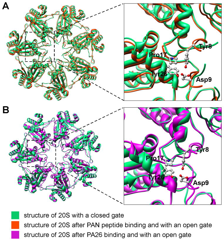
Substrates enter the cylindrical 20S proteasome through a gated channel that is regulated by the ATPases in the 19S regulatory particle in eukaryotes or the homologous PAN ATPase complex in archaea. These ATPases contain a conserved C-terminal hydrophobic-tyrosine-X (HbYX) motif that triggers gate opening upon ATP binding. Using cryo-electron microscopy, we identified the sites in the archaeal 20S where PAN's C-terminal residues bind and determined the structures of the gate in its closed and open forms. Peptides containing the HbYX motif bind to 20S in the pockets between neighboring alpha subunits where they interact with conserved residues required for gate opening. This interaction induces a rotation in the alpha subunits and displacement of a reverse-turn loop that stabilizes the open-gate conformation. This mechanism differs from that of PA26/28, which lacks the HbYX motif and does not cause alpha subunit rotation. These findings demonstrated how the ATPases' C termini function to facilitate substrate entry.


Figures

Figure 1
Figure 1
Difference map reveals the binding sites of the peptides to 20S. A and B: Top and side (cut in half and looking from inside-out) views of 3D density map of 20S (yellow) with the atomic structure (pdb: 1pma, green ribbon diagram) docked. Superimposed are difference densities (blue, 6σ, where σ is rms deviation from mean) between the 20S-AHLDVLYA and the 20S. C: Top and side views of two adjacent α-subunits (light green) with the difference densities that indicate the binding sites of the peptides. The side chain of Lys66 is shown. D: Structure of the β-ring (dark green) superimposed with the difference densities that correspond to peptides bound to the proteolytic sites. The side chains of the active site Thr1 are shown.
Figure 2
Figure 2
The closed and the open gate of 20S. A and B: Density maps of the closed gate (A, yellow) and the open gate (B, blue), with the structure of the docked α-subunits (ribbon diagram). C and D: Densities of an individual α-subunit boxed out from A and B. The symmetry axes are marked. Beyond Thr13 (pointed by arrowheads) the structures are modeled (magenta) to fit the densities of both the closed and open gate conformations. The dashed line in C marked a possible way the remaining residues in the N-terminus may extend further into the pore.
Figure 3
Figure 3
Conformational change of 20S α-ring induced by the gate-opening peptides. A: Top view of superimposed density maps of 20S (gold mesh) and 20S-AHLDVLYA complex (blue transparent surface). B: Top view of the 20S-AHLDVLYA density map (blue) with the structure of the open-gate 20S docked. C: Top view of the 20S density map (gold) with the structure of the closed-gate 20S docked. D: Superimposed structures of 20S with the open and the closed gate.
Figure 4
Figure 4
Interactions of the gate-opening peptides with the 20S. A: Top view of the pocket between adjacent α-subunits with (left) and without (right) peptide bound. B: side view of the same pocket (90° rotated) and the density slab within the dashed line in A with (left) and without (right) peptide bound. Residues that may be involved in peptide binding are marked. Notice that the density pointed by the arrowhead (left) is not present in the density map without the bound peptide (right).
Figure 5
Figure 5
Abilities of three different activators to stimulate the gate opening of 20S mutants. 0.2μg of 20S proteasome (wild type or various Leu81/Val82 mutants) and LFP are incubated with (A) PAN in a molar ratio of 10:1 of 20S, (B) 250 μM of peptide HLDVLYR, and (C) PA26 in a molar ratio of 30:1 of 20S. The stimulation of gate opening was measured by the increase of LFP hydrolysis over wild type 20S without any activator. The values are means ± standard deviations from at least three independent experiments.
Figure 6
Figure 6
Mechanism of gate opening in the 20S induced by PAN and PA26 complexes. A Left: overlay of the α-ring structures, before and after the binding of the gate-opening peptides. Right: enlarged view of the dashed area from the left. Pro17 is shifted because of the rotation in α-subunit. B: Left: Overlay of the structure of the α-ring, before and after PA26 binding. Right: enlarged view of the dashed area. Pro17 is shifted by the activation loop of PA26 (not shown). Notice that Pro17 in red and magenta structures are in a similar position.

References

    1. Benaroudj N, Goldberg AL. PAN, the proteasome-activating nucleotidase from archaebacteria, is a protein-unfolding molecular chaperone. Nat Cell Biol. 2000;2:833–839. - PubMed
    1. Benaroudj N, Zwickl P, Seemuller E, Baumeister W, Goldberg AL. ATP hydrolysis by the proteasome regulatory complex PAN serves multiple functions in protein degradation. Mol Cell. 2003;11:69–78. - PubMed
    1. Coux O, Tanaka K, Goldberg AL. Structure and functions of the 20S and 26S proteasomes. Annu Rev Biochem. 1996;65:801–847. - PubMed
    1. Crowther RA, Henderson R, Smith JM. MRC image processing programs. J Struct Biol. 1996;116:9–16. - PubMed
    1. DeLaBarre B, Brunger AT. Nucleotide dependent motion and mechanism of action of p97/VCP. J Mol Biol. 2005;347:437–452. - PubMed

Publication types

MeSH terms

LinkOut - more resources