Review
. 2008 Sep;1786(1):15-23.
doi: 10.1016/j.bbcan.2008.04.002. Epub 2008 Apr 22.

P53, cyclin-dependent kinase and abnormal amplification of centrosomes

Affiliations
Review

P53, cyclin-dependent kinase and abnormal amplification of centrosomes

Kenji Fukasawa. Biochim Biophys Acta. 2008 Sep.

Abstract

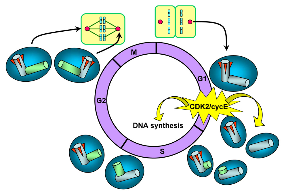
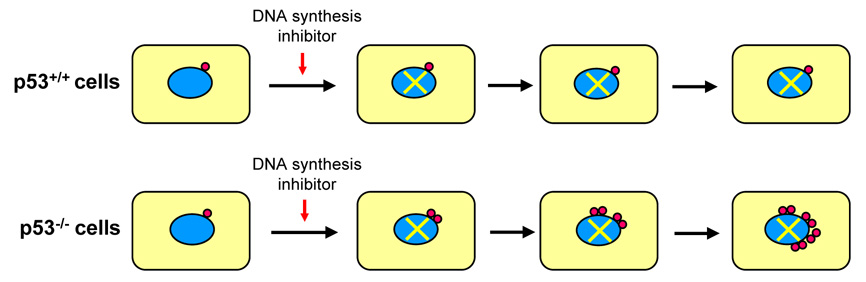
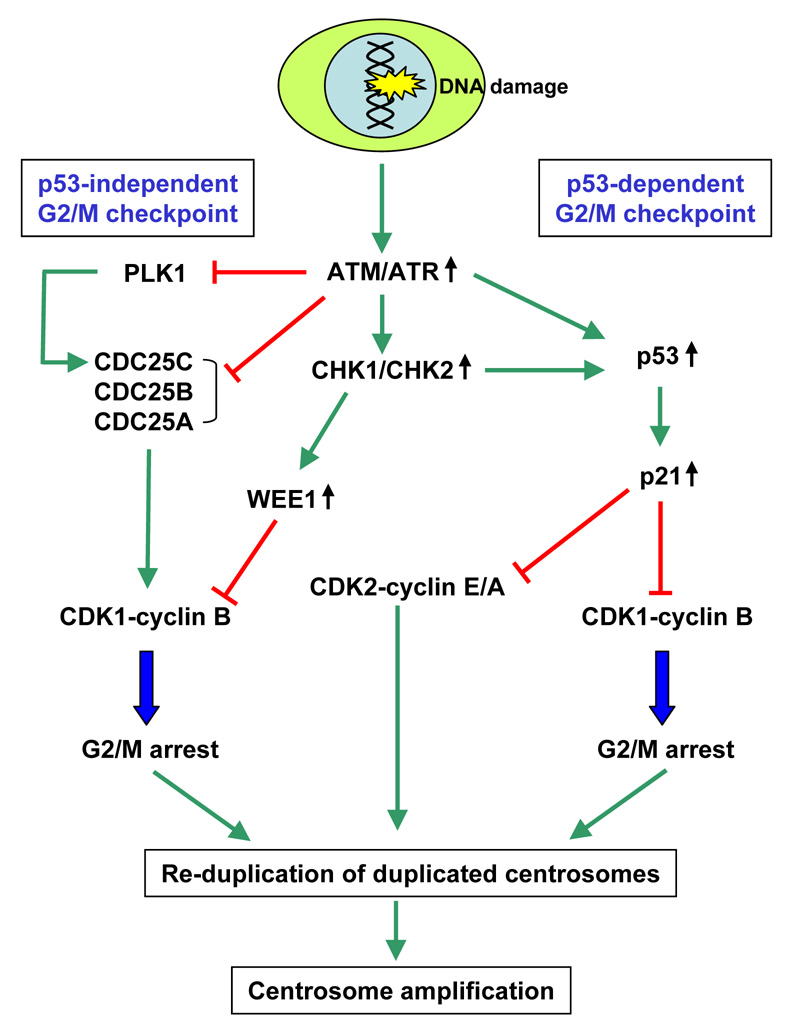
Centrosomes play a critical role in formation of bipolar mitotic spindles, an essential event for accurate chromosome segregation into daughter cells. Numeral abnormalities of centrosomes (centrosome amplification) occur frequently in cancers, and are considered to be the major cause of chromosome instability, which accelerates acquisition of malignant phenotypes during tumor progression. Loss or mutational inactivation of p53 tumor suppressor protein, one of the most common mutations found in cancers, results in a high frequency of centrosome amplification in part via allowing the activation of the cyclin-dependent kinase (CDK) 2-cyclin E (as well as CDK2-cyclin A) which is a key factor for the initiation of centrosome duplication. In this review, the role of centrosome amplification in tumor progression, and mechanistic view of how centrosomes are amplified in cells through focusing on loss of p53 and aberrant activities of CDK2-cyclins will be discussed.


Figures

Figure 1
Figure 1. Structure and function of centrosomes
(a) The basic structure of the centrosome. (b) During mitosis, two centrosomes become the spindle poles, directing the formation of bipolar mitotic spindles.
Figure 2
Figure 2. Mitotic defects associated with numeral abnormalities of centrosomes
In normal mitosis, two centrosomes direct the formation of bipolar mitotic spindles (a). In the presence of amplified centrosomes, cells frequently form multiple (>2) spindle poles. (b) Tripolar spindles can undergo cytokinesis, and some daughter cells are viable, yet suffer severe aneuploidy. (c) Cells with spindles with >3 poles fail to undergo cytokinesis in most cases, becoming either bi-nucleated or large mono-nucleated cells. Because of the p53-dependent checkpoint response to cytokinesis failure, the cells become arrested in the presence of p53, and eventually undergo cell death. In contrast, in the absence of p53, cells continue to cycle, and many of them become very large multi(>2)-nucleated cells and often undergo senescence-like arrest and cell death. However, some cells escape from continuous cytokinesis block, and become polyploid cells.
Figure 3
Figure 3. Pseudo-bipolar spindles and the risk of chromosome segregation errors
Amplified centrosomes frequently form pseudo-bipolar spindles by positioning on a bipolar axis (centrosome clustering), resulting in mitotic spindles which structurally resemble the true bipolar spindles organized by two centrosomes. Cells with pseudo-bipolar spindles undergo normal cytokinesis without any chromosome segregation errors (a). However, one or a few centrosomes often fail to position on the bipolar axis. Those mal-positioned centrosomes are still functionally intact, nucleating microtubules which capture chromosomes. Depending on whether those particular chromosomes are segregated into one or the other daughter cell, aneuploid daughter cells can be generated (b).
Figure 4
Figure 4. Karyotypic convergence during tumor progression
The early stage tumors are often karyotypically heterogeneous, while the late stage tumors are karyotypically homogenous. The karyotypic convergence theory provides the mechanism underlying this phenomenon. A cell acquires a mutation that renders growth advantage over the others, and clonally expands (a). Among this particular population of cells, one or a few cells acquire the mutations that destabilize chromosomes (CIN phenotype, i.e., centrosome amplification) (b). Because of the CIN phenotype, the population gradually becomes karyotypically heterogeneous (c), representing the early stage tumor. A cell within this population eventually acquires the favorable chromosome composition that renders maximal growth/carcinogenic potentials (d), and clonally expands (f). For these cells, maintenance of this particular chromosome composition becomes the priority, selecting the cell(s) that have acquired the mutation(s) that either counteract or eliminate the cause of CIN (g). Such a cell will eventually dominate the population by retaining the favorable karyotypic composition, and thus the population becomes karyotypically homogenous, representing late stage tumors.
Figure 5
Figure 5. The centrosome duplication cycle
CDK2/cyclin E, a known inducer of S-phase entry, is also a key initiator of centrosome duplication. Thus, the late G1 specific activation of CDK2/cyclin E is at least in part responsible for the coordinated initiation of centrosome and DNA duplication. Centrosome duplication begins with the physical splitting of the paired centrioles, followed by formation of procentrioles near the proximal end of each pre-existing centriole. During S and G2, procentrioles elongate, and two centrosomes progressively recruit PCM, an event known as centrosome maturation. In late G2, the daughter centriole of the parental pair acquires sub-distal appendages (red wedges), and two mature centrosomes are generated. At late G2 prior to mitosis, two duplicated centrosomes separate and migrate to opposite ends of the cell. During mitosis, duplicated centrosomes form spindle poles to direct the formation of bipolar mitotic spindles. Upon cytokinesis, each daughter cell receives one centrosome along with one half of the duplicated DNA.
Figure 6
Figure 6. Centrosome re-duplication in cells arrested by exposure to DNA synthesis inhibitors in the absence of p53
When cells are exposed to DNA synthesis inhibitors such as hydroxyurea, aphidicolin and mimosine, cells become arrested at the G1/S transition and in S-phase in a p53-independent manner. In the presence of the functionally intact p53, p53 is stabilized/activated in response to the stress associated with prolonged arrest by the ARF-dependent mechanism, leading to up-regulation of p21, which in turn continuously inhibits CDK2, hence blocking centrosome duplication (a). In contrast, if p53 is either lost or mutated, CDK2 activity is unchecked and activated, which triggers centrosome re-duplication in the absence of DNA synthesis, resulting in generation of amplified centrosomes (b).
Figure 7
Figure 7. Centrosome amplification in cells arrested by the G2/M checkpoint in response to DNA damage occurs efficiently in the absence of p53
The G2/M checkpoints in response to DNA damage are exerted through the p53-dependent and p53-independent pathways, both of which target CDK1-cyclin B. Both p53-dependent and - independent G2/M checkpoint responses are triggered by activation of ATM/ATR and CHK1/CHK2 kinases. In the p53-independent pathway, CHK1 and CHK2 phosphorylate and inhibit CDC25A, B and C. CHK1 also inhibits the activity of PLK1, which is known to activate CDC25C. CHK1 also appears to suppress CDC25B by blocking the access of CDC25B to centrosomally localized CDK1-cyclin B (initial activation of CDK1-cyclin B is thought to occur at centrosomes) [85, 86]. CHK1 also phosphorylates and up-regulates the activity of WEE1 kinase that catalyzes the inhibitory phosphorylation of CDK1. All these events block the activation of CDK1-cyclin B in a concerted fashion, resulting in late G2 arrest. In the p53-dependent pathway, ATM/ATR and CHK1/CHK2 phosphorylate and stabilize p53, which in turn up-regulates p21. p21 then suppresses the activity of CDK1-cyclin B, leading to late G2 arrest. This G2 arrest provides time for centrosomes to regain duplication competency and re-duplicate. However, CDK2 activity is still required for centrosomes to re-duplicate. In late G2 phase, high levels of already active form of CDK2 (mostly complexed with cyclin A) are present. In cells arrested by the G2/M checkpoint responses, those CDK2 kinases remain active even efficient inhibition of CDC25s. However, if the intact p53-dependent pathway is present, centrosome re-duplication is blocked by the p21-mediated suppression of the CDK2 activity. In contrast, in the absence of the p53-dependent pathway, centrosomes re-duplication is continuously triggered by active CDK2-cyclin E/A.

Similar articles

Cited by

References

    1. Bornens M. Centrosome composition and microtubule anchoring mechanisms. Curr. Opin. Cell Biol. 2002;14:25–34. - PubMed
    1. Sattler CA, Sawada N, Sattler GL, Pitot HC. Electron microscopic and time lapse studies of mitosis in cultured rat hepatocytes. Hepatology. 1988;8:1540–1549. - PubMed
    1. Uetake Y, Sluder G. Cell cycle progression after cleavage failure: mammalian somatic cells do not possess a "tetraploidy checkpoint". J. Cell Biol. 2004;165:609–615. - PMC - PubMed
    1. Fukasawa K. Centrosome amplification, chromosome instability and cancer development. Cancer Lett. 2005;230:6–19. - PubMed
    1. Levine DS, Sanchez CA, Rabinovitch PS, Reid BJ. Formation of the tetraploid intermediate is associated with the development of cells with more than four centrioles in the elastase-simian virus 40 tumor antigen transgenic mouse model of pancreatic cancer. Proc. Natl. Acad. Sci. USA. 1991;88:6427–6431. - PMC - PubMed

Publication types

MeSH terms