. 2008 May;10(5):481-8.
doi: 10.1593/neo.08178.

Cathepsin B mediates the pH-dependent proinvasive activity of tumor-shed microvesicles

Affiliations

Cathepsin B mediates the pH-dependent proinvasive activity of tumor-shed microvesicles

Ilaria Giusti et al. Neoplasia. 2008 May.

Abstract

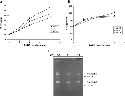
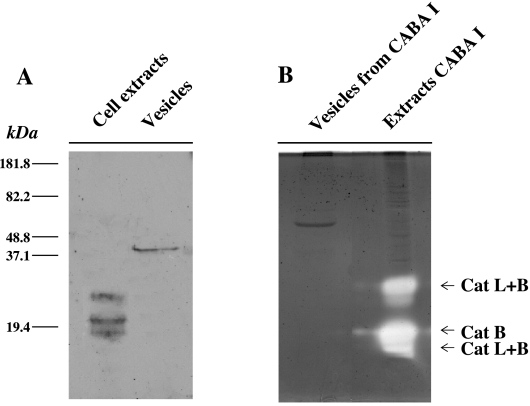
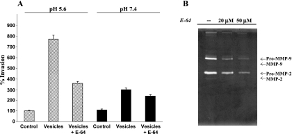
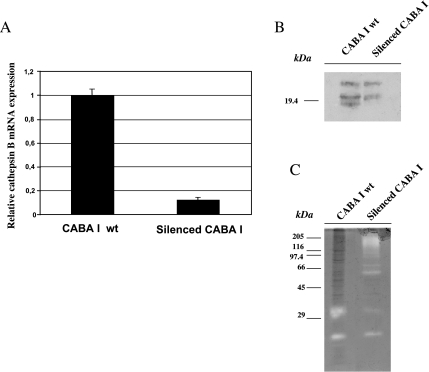
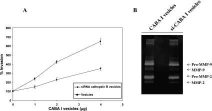
Vesicles shed by cancer cells are known to mediate several tumor-host interactions. Tumor microenvironment may, in turn, influence the release and the activity of tumor-shed microvesicles. In this study, we investigated the molecular mediators of the pH-dependent proinvasive activity of tumor-shed vesicles. Gelatinase zymography showed increased microvesicle activity of matrix metalloproteinases 9 and 2 as a result of acid exposure (pH 5.6) compared to pH 7.4. Thus, we reasoned that the cysteine protease cathepsin B might play a role in mediating the pH-dependent activation of gelatinases. Cathepsin B expression in tumor-shed microvesicles was confirmed by Western blot analysis and zymography. The activity of vesicle-associated cathepsin B measured using Z-Arg-Arg-pNA as substrate was significantly increased at acidic pH values. Inhibition of protease activity by the cysteine protease inhibitor, E-64, and treatment of ovarian cancer cells with small interfering RNA against cathepsin B suppressed the ability of tumor-shed microvesicles to stimulate both gelatinase activation and the invasiveness of endothelial cells observed at low pH values. We conclude that microvesicle shedding is a major secretory pathway for cathepsin B release from tumor cells. Hence, the acidic microenvironment found in most solid tumors may contribute to cathepsin B-mediated proinvasive capabilities of tumor-shed vesicles.


Figures

Figure 1
Figure 1
Effect of tumor-shed vesicles on endothelial cell motility and invasiveness. Human umbilical vein endothelial cell invasiveness (A) and motility (B) were tested in the Boyden chamber. Vesicles shed by human ovarian carcinoma cells CABA I were used as external stimuli. Data (mean ± SD of three independent experiments) represent the number of cells that had migrated in five high-power fields expressed in percentage, counting untreated cells as 100%, P < .05. (C) Effect of acidic pH on vesicle-associated gelatinases.
Figure 2
Figure 2
Presence of cathepsin B in CABA I-shed vesicles. (A) Western blot analysis of cell extracts and vesicle-associated cathepsin B. (B) Cysteine zymography of extracts and vesicle-associated cathepsins B and L. The identification of cathepsin B was confirmed through the inhibition of cathepsin L (not shown).
Figure 3
Figure 3
Effect of cathepsin B inhibition. (A) Vesicles shed from CABA I were isolated and tested for their ability to stimulate HUVEC invasiveness at pH 5.6 and 7.4, either with or without the cysteine proteinase inhibitor E-64 (50 µM). Data (mean ± SD of three independent experiments) represent the number of cells that had migrated in five high-power fields expressed in percentage, counting untreated cells as 100%, P < .05. (B) Zymographic analysis of vesicle-associated gelatinases incubated with inhibitor E-64 0 to 50 µM.
Figure 4
Figure 4
Effectiveness of cathepsin B siRNA silencing in CABA I cells. (A) RT-PCR analysis of parental control and silenced CABA I cells. (B) Western blot analysis of cathepsin B in cellular extracts. (C) Cysteine zymography of cathepsins B and L in cellular extracts.
Figure 5
Figure 5
Effect of cathepsin B gene silencing. (A) Vesicles shed from CABA I cells, either parental control and silenced, were isolated and tested for their ability to stimulate HUVEC invasiveness at pH 5.6. Data (mean ± SD of three independent experiments) represent the number of cells that had migrated in five high-power fields expressed in percentage, counting untreated cells as 100%, P < .05. (B) Zymographic analysis of vesicle-associated gelatinases.

References

    1. Yan L, Shahied L, Tang Y, Kesavan P, Nakada MT. Tumor-host interactions. Preclinica. 2004;2:422–426.
    1. Zigrino P, Loffek S, Mauch C. Tumor-stroma interactions: their role in the control of tumor cell invasion. Biochimie. 2005;87:321–328. - PubMed
    1. Laconi E. The evolving concept of tumor microenvironments. Bioessays. 2007;29:738–744. - PubMed
    1. Taraboletti G, D'Ascenzo S, Borsotti P, Giavazzi R, Pavan A, Dolo V. Shedding of the matrix metalloproteinases MMP-2, MMP-9, and MT1-MMP as membrane vesicle-associated components by endothelial cells. Am J Pathol. 2002;160:673–680. - PMC - PubMed
    1. Dolo V, Ginestra A, Ghersi G, Nagase H, Vittorelli ML. Human breast carcinoma cells cultured in the presence of serum shed membrane vesicles rich in gelatinolytic activities. J Submicrosc Cytol Pathol. 1994;26:173–810. - PubMed

Publication types

MeSH terms

LinkOut - more resources