Review
. 2008 May;8(4):381-9.
doi: 10.2174/187152008784220357.

Tyrosyl-DNA phosphodiesterase as a target for anticancer therapy

Affiliations
Review

Tyrosyl-DNA phosphodiesterase as a target for anticancer therapy

Thomas S Dexheimer et al. Anticancer Agents Med Chem. 2008 May.

Abstract

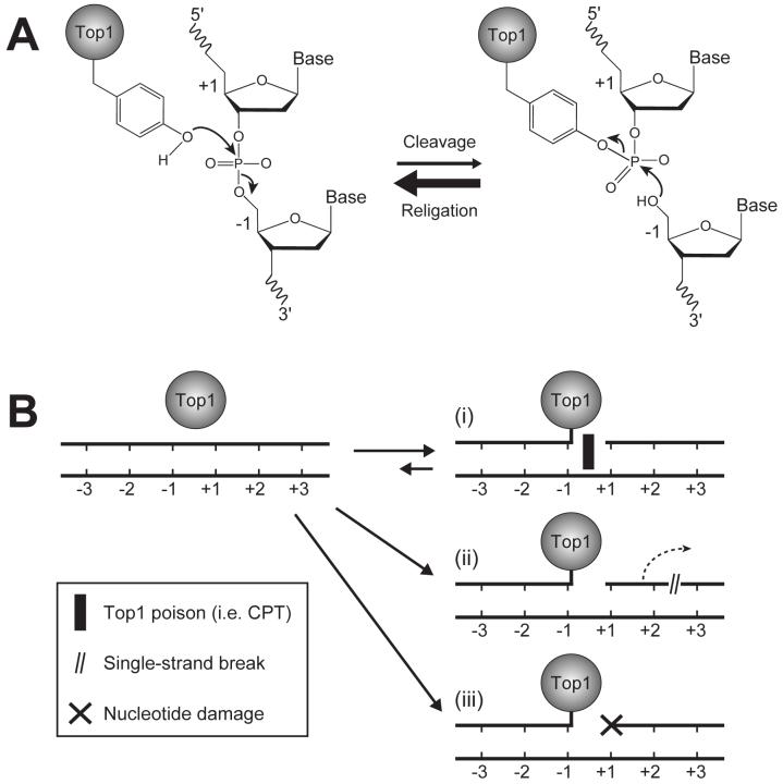
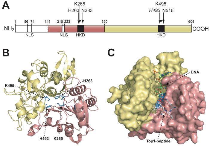
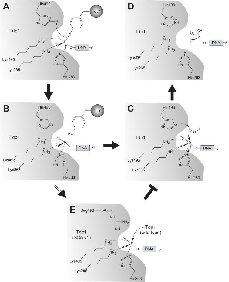
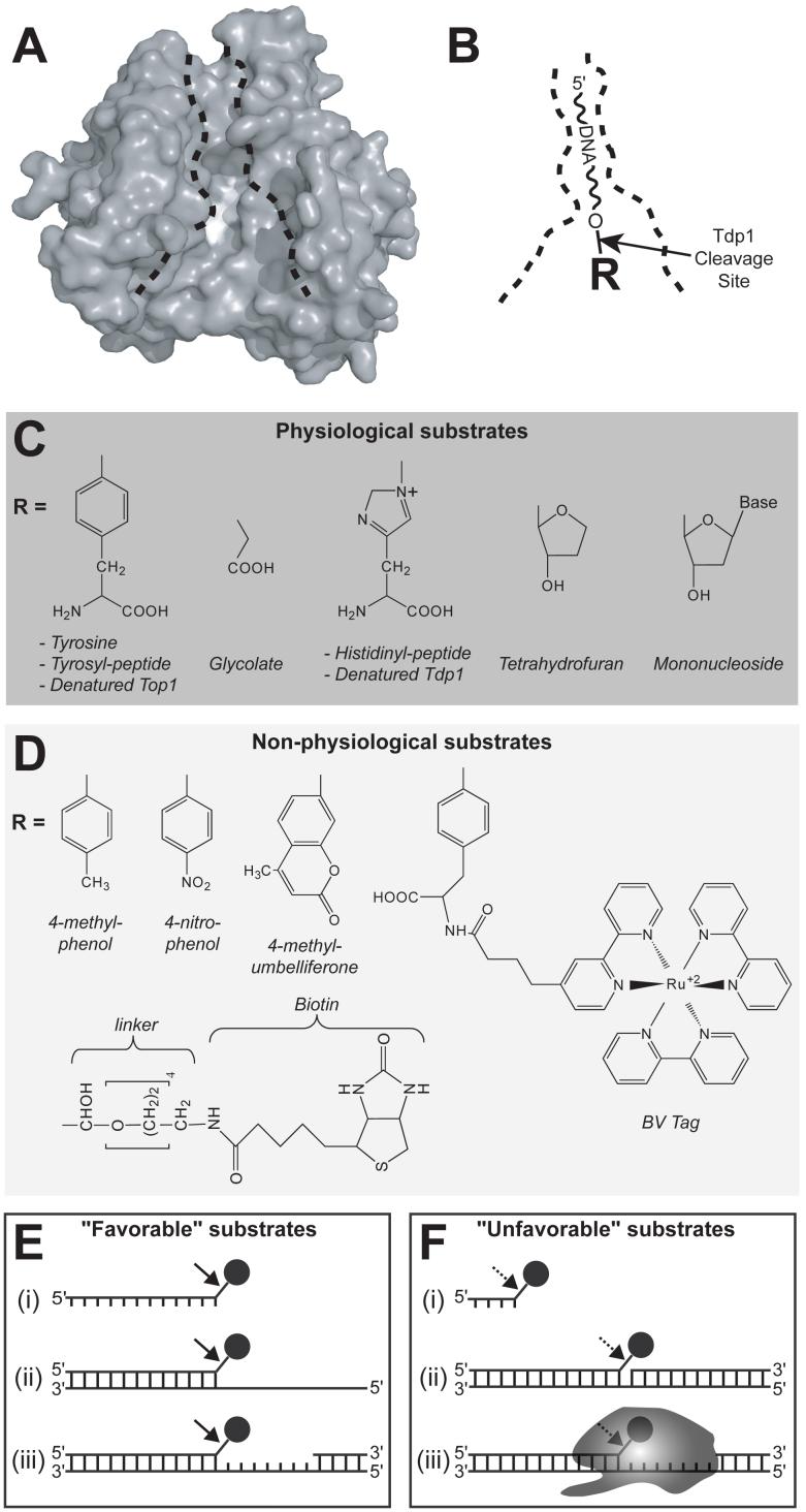
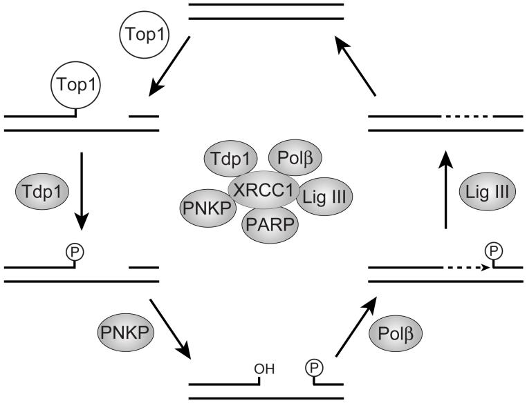
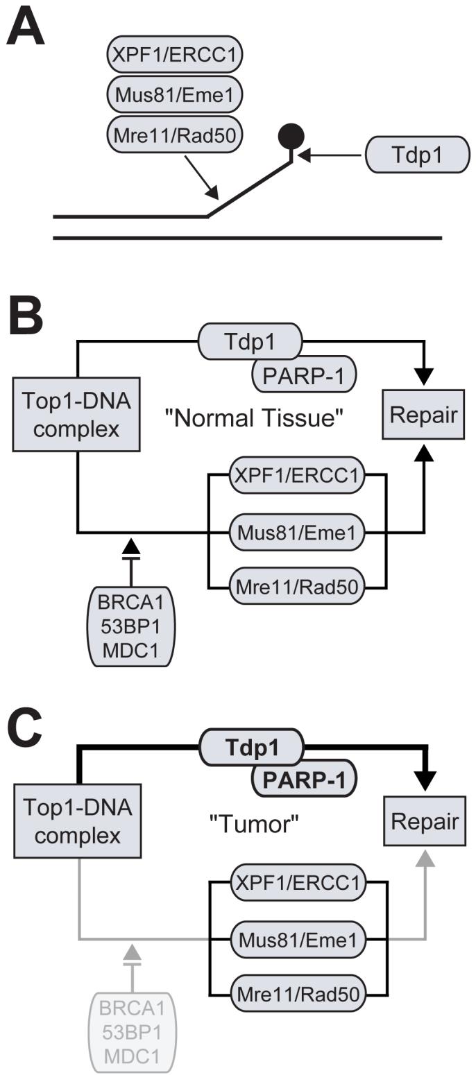
Tyrosyl-DNA phosphodiesterase 1 (Tdp1) is a recently discovered enzyme that catalyzes the hydrolysis of 3'-phosphotyrosyl bonds. Such linkages form in vivo following the DNA processing activity of topoisomerase I (Top1). For this reason, Tdp1 has been implicated in the repair of irreversible Top1-DNA covalent complexes, which can be generated by either exogenous or endogenous factors. Tdp1 has been regarded as a potential therapeutic co-target of Top1 in that it seemingly counteracts the effects of Top1 inhibitors, such as camptothecin and its clinically used derivatives. Thus, by reducing the repair of Top1-DNA lesions, Tdp1 inhibitors have the potential to augment the anticancer activity of Top1 inhibitors provided there is a presence of genetic abnormalities related to DNA checkpoint and repair pathways. Human Tdp1 can also hydrolyze other 3'-end DNA alterations including 3'-phosphoglycolates and 3'-abasic sites indicating it may function as a general 3'-DNA phosphodiesterase and repair enzyme. The importance of Tdp1 in humans is highlighted by the observation that a recessive mutation in the human TDP1 gene is responsible for the inherited disorder, spinocerebellar ataxia with axonal neuropathy (SCAN1). This review provides a summary of the biochemical and cellular processes performed by Tdp1 as well as the rationale behind the development of Tdp1 inhibitors for anticancer therapy.


Figures

Fig. (1)
Fig. (1)
A) Human Top1-mediated DNA cleavage and religation mechanisms. Tyr723 is the active site tyrosine involved in the transesterification reaction. The bases flanking the Top1 cleavage site are referred to as -1 and +1 for the bases at the 3′ and 5′ DNA termini, respectively. B) Representative circumstances resulting in the formation of trapped Top1 cleavage complexes (i.e. (i) Top1 inhibitors or preexisting DNA lesions, such as (ii) DNA strand breaks or (iii) nucleotide base damage).
Fig. (2)
Fig. (2)
A) Schematic representation of the human Tdp1 domain structure. The N-terminal and C-terminal domains correspond to residues 1-350 and 351-608, respectively. Positions of the “HKN” motifs and NLS (nuclear localization signals) motifs are shown in black and white, respectively. Arrows indicate active site residues. B) Ribbon diagram of Tdp1 structure (Δ148). The domain colors correspond to those shown in (A). The active site residues (H263, K265, H493, and K495) are shown as stick structures in blue (Figure modified from [40]). C) Structure of the Tdp1-vanadate-peptide-DNA complex (1NOP). The orientation and domain colors of Tdp1 are identical as in (B). Tdp1 is shown as a molecular surface and the vanadate-peptide-DNA substrate mimic is shown as ball-and-stick structures with the DNA colored in green, the vanadate colored in red, and the Top1-derived peptide colored in white (Figure modified from [41]).
Fig. (3)
Fig. (3)
Proposed reaction mechanism for human Tdp1. A) The first step of the reaction involves the nucleophilic attack of the phosphodiester-containing substrate by the imidazole Nε2 atom of His263. H493 donates a proton to the tyrosyl moiety of the leaving group. The phosphohistidine intermediate is shown in (B). Interactions between the non-bridging oxygens and the active site lysine residues (Lys265 and Lys495) serve to stabilize the transition intermediate. C) The next step of the reaction involves second a nucleophilic attack by a water molecule activated by H493, generating final 3′-phosphate product and free Tdp1 (D) (A-D modified from [40]). (E) The SCAN-1 mutations (H493R) leads to an accumulation of the Tdp1-DNA intermediate and a defect in Tdp1 turnover rate.
Fig. (4)
Fig. (4)
Outline of the substrate binding pocket and the different substrates processed by Tdp1. A) Molecular surface of Tdp1 in an orientation identical to that shown in Fig. 2C. The active site region is shown in white. The black dotted lines define the substrate binding pocket (Figure modified from [40]). B) Substrate orientation in the Tdp1 binding pocket. The R-group refers to the structures shown in (C) and (D). The arrow indicates the Tdp1 cleavage site. C) and D) illustrate the physiological and non-physiological substrates of Tdp1 (Figure modified and updated from [61]). E) and F) show both favorable and unfavorable the Tdp1 substrates. The black circle represents a 3′-end phosphotyrosine. E) (i) long single-strand, (ii) tailed duplex, (iii) gapped duplex. F) (i) short single-strand, (ii) nicked duplex, (iii) full-length Top1.
Fig. (5)
Fig. (5)
Proposed repair of Top1 cleavage complexes by the XRCC-1-dependent pathway. The XRCC1 complex including the associated repair enzymes is shown in the center of the figure. Tdp1 hydrolyzes the Top1 phosphotyrosyl bond. PNKP hydrolyzes the resulting 3′-phosphate and phosphorylates the 5′-hydroxyl. DNA polymerase β fills the gap and DNA ligase III reseals the DNA. PARP-1 may play a role in the initial recognition of the Top1-mediated DNA damage. (Figure modified from [8, 19, 76])
Fig. (6)
Fig. (6)
Redundant pathways involved the repair of Top1 cleavage complexes in mammalian cells. A) The processing of a Top1 cleavage complex by Tdp1 and three different structure-specific 3′-endonucleases. B) Schematic representation of the Tdp1-dependent pathway and checkpoint-dependent 3′-endonuclease pathway, which together are implicated in the repair of Top1 cleavage complexes. B) DNA repair and checkpoint deficiencies, such as in cancer cells, results in enhanced dependency for the repair of Top1 cleavage complexes via the Tdp1-dependent pathway.
Fig. (7)
Fig. (7)
Chemical structures of Tdp1 inhibitors reported to date.

References

    1. Champoux JJ. Annu. Rev. Biochem. 2001;70:369. - PubMed
    1. Keck JL, Berger JM. Nat. Struct. Biol. 1999;6:900. - PubMed
    1. Pommier Y, Pourquier P, Fan Y, Strumberg D. Biochim. Biophys. Acta. 1998;1400:83. - PubMed
    1. Wang JC. Nat. Rev. Mol. Cell Biol. 2002;3:430. - PubMed
    1. Hsiang YH, Hertzberg R, Hecht S, Liu LF. J. Biol. Chem. 1985;260:14873. - PubMed

Substances