. 2008 May 12:5:14.
doi: 10.1186/1742-2094-5-14.

The synthetic triterpenoid CDDO-methyl ester modulates microglial activities, inhibits TNF production, and provides dopaminergic neuroprotection

Affiliations

The synthetic triterpenoid CDDO-methyl ester modulates microglial activities, inhibits TNF production, and provides dopaminergic neuroprotection

Thi A Tran et al. J Neuroinflammation. .

Abstract

Background: Recent animal and human studies implicate chronic activation of microglia in the progressive loss of CNS neurons. The inflammatory mechanisms that have neurotoxic effects and contribute to neurodegeneration need to be elucidated and specifically targeted without interfering with the neuroprotective effects of glial activities. Synthetic triterpenoid analogs of oleanolic acid, such as methyl-2-cyano-3,12-dioxooleana-1,9-dien-28-oate (CDDO-Me, RTA 402) have potent anti-proliferative and differentiating effects on tumor cells, and anti-inflammatory activities on activated macrophages. We hypothesized that CDDO-Me may be able to suppress neurotoxic microglial activities while enhancing those that promote neuronal survival. Therefore, the aims of our study were to identify specific microglial activities modulated by CDDO-Me in vitro, and to determine the extent to which this modulation affords neuroprotection against inflammatory stimuli.

Methods: We tested the synthetic triterpenoid methyl-2-cyano-3,12-dioxooleana-1,9-dien-28-oate (CDDO-Me, RTA 402) in various in vitro assays using the murine BV2 microglia cell line, mouse primary microglia, or mouse primary peritoneal macrophages to investigate its effects on proliferation, inflammatory gene expression, cytokine secretion, and phagocytosis. The antioxidant and neuroprotective effects of CDDO-Me were also investigated in primary neuron/glia cultures from rat basal forebrain or ventral midbrain.

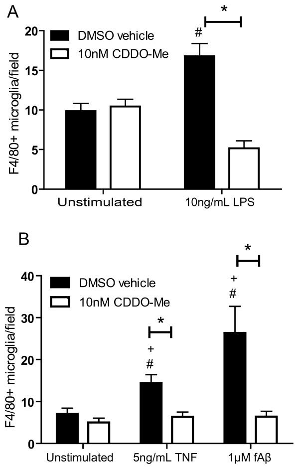
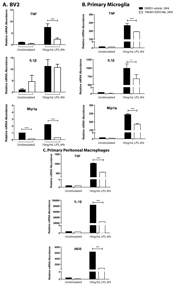
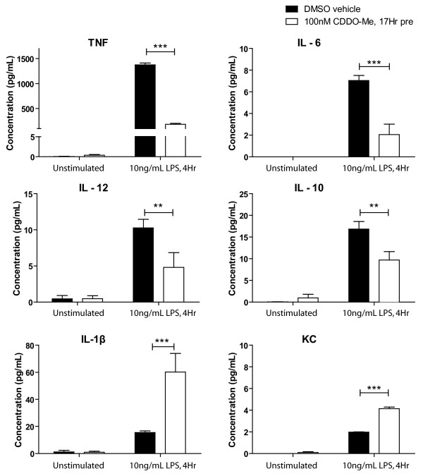
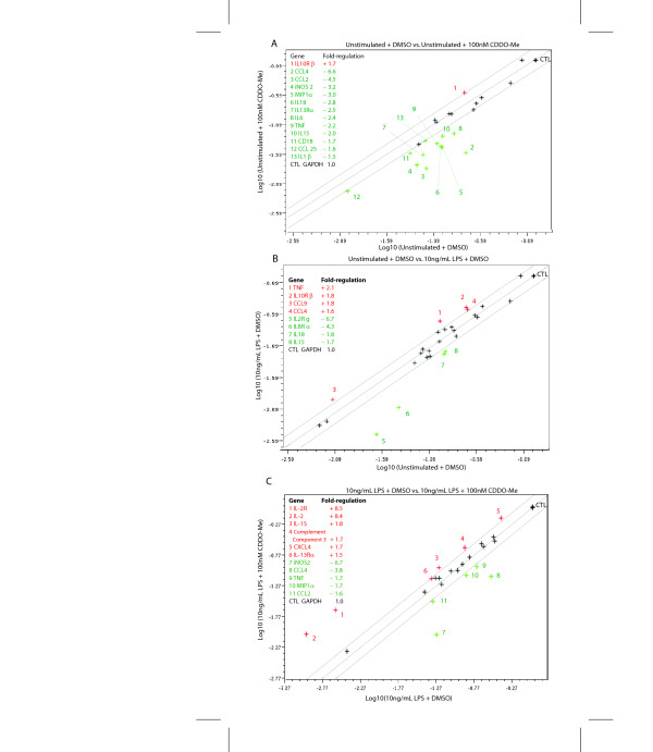
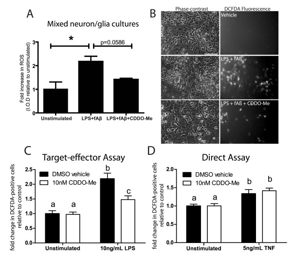
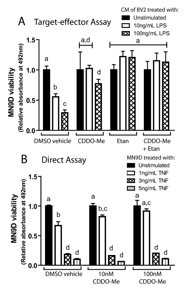
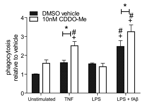
Results: We found that at low nanomolar concentrations, treatment of rat primary mesencephalon neuron/glia cultures with CDDO-Me resulted in attenuated LPS-, TNF- or fibrillar amyloid beta 1-42 (A beta 1-42) peptide-induced increases in reactive microglia and inflammatory gene expression without an overall effect on cell viability. In functional assays CDDO-Me blocked death in the dopaminergic neuron-like cell line MN9D induced by conditioned media (CM) of LPS-stimulated BV2 microglia, but did not block cell death induced by addition of TNF to MN9D cells, suggesting that dopaminergic neuroprotection by CDDO-Me involved inhibition of microglial-derived cytokine production and not direct inhibition of TNF-dependent pro-apoptotic pathways. Multiplexed immunoassays of CM from LPS-stimulated microglia confirmed that CDDO-Me-treated BV2 cells produced decreased levels of specific subsets of cytokines, in particular TNF. Lastly, CDDO-Me enhanced phagocytic activity of BV2 cells in a stimulus-specific manner but inhibited generation of reactive oxygen species (ROS) in mixed neuron/glia basal forebrain cultures and dopaminergic cells.

Conclusion: The neuroimmune modulatory properties of CDDO-Me indicate that this potent antioxidant and anti-inflammatory compound may have therapeutic potential to modify the course of neurodegenerative diseases characterized by chronic neuroinflammation and amyloid deposition. The extent to which synthetic triterpenoids afford therapeutic benefit in animal models of Parkinson's and Alzheimer's disease deserves further investigation.


Figures

Figure 1
Figure 1
CDDO-Me attenuates LPS-, TNF-, and fibrillar Aβ42-induced primary microglia activation. Rat embryonic ventral mesencephalon primary cultures were treated at day 5 in vitro with indicated compounds (LPS 10 ng/mL, TNF 5 ng/mL, fAβ 1 μM, CDDO-Me 10 nM). Cells were fixed at 2 days post-treatment and stained with an antibody against the microglial activation marker F4/80. Values are expressed as mean number of F4/80-positive microglia per field ± S.E.M.. Values were analyzed by two-way ANOVA followed by Tukey post hoc test, * denotes CDDO-Me is significantly different from its DMSO vehicle for a given treatment; # denotes significant difference from the DMSO vehicle in unstimulated cells; + denotes significant difference from CDDO-Me in stimulated cells; all symbols at p < 0.05.
Figure 2
Figure 2
CDDO-Me downregulates expression of mRNAs for pro-inflammatory genes. (A) BV2 microglia were pre-treated with 10 nM CDDO-Me for 17 hr before a 4-hr stimulation as indicated. (B) Microglia from post-natal day 2 (P2) wild-type mice were pre-treated with 100 nM CDDO-Me for 24 hr before a 4 hr stimulation as indicated. (C) Peripheral macrophages from adult wild-type mice were pre-treated with 100 nM CDDO-Me for 24 hr before a 4-hr stimulation as indicated. Values represent mean ± S.E.M. and are expressed relative to vehicle-treated, vehicle-stimulated conditions. Values were analyzed by two-way ANOVA followed by Bonferroni's post hoc. * denotes significance at p < 0.05, *** denotes significance at p < 0.001.
Figure 3
Figure 3
CDDO-Me suppresses production of cytokines with known neurotoxic effects on dopaminergic cultures. BV2 microglia were pre-treated with 100 nM CDDO-Me or DMSO vehicle for 17 hrs before a 4-hr stimulation with LPS (10 ng/mL). Inflammatory cytokine and chemokine production were measured from CM using a 7-plex inflammatory cytokine profile immunoassay from Meso-Scale Discovery on an MSD 2400 plate reader. Values represent mean cytokine production ± SEM. Values were analyzed by two way ANOVA followed by Bonferroni's post hoc. ** denotes significance at p < 0.01, *** denotes significance at p < 0.001.
Figure 4
Figure 4
CDDO-Me suppresses basal and inflammation-induced gene expression. BV2 microglia cultures were pre-incubated with 100 nM CDDO-Me for 17 hrs before a 4-hr stimulation with 10 ng/mL LPS in serum-free medium. (A) Scatterplot of baseline inflammatory gene expression in unstimulated BV2 cells in the presence of DMSO vehicle (0.1%) versus unstimulated cells in the presence of 100 nM CDDO-Me. (B) Unstimulated cells in the presence of DMSO vehicle versus cells stimulated with LPS (10 ng/mL) in the presence of DMSO vehicle, or (C) cells stimulated with LPS in presence of DMSO vehicle versus cells stimulated with LPS in the presence of 10 nM CDDO-Me plus. Data analyses were performed using the Scatter Plot data analysis tool in the SuperArray GEArray Analysis Suite online (See Methods). Genes shown with a red plus (+) sign were up-regulated and genes shown with a green minus (-) sign were down-regulated in the treatment condition, which is plotted on the Y-axis relative to the control condition plotted on the X-axis.
Figure 5
Figure 5
CDDO-Me inhibits intracellular ROS accumulation induced by LPS and fibrillar Aβ42 in neuronal cultures from E14 rat basal forebrain cholinergic mixed neuron-glia cultures (BFCs) and in dopaminergic cells. (A) Fold-change in intracellular ROS. Integrated optical density was measured from digital images of treated BFCs. Results are expressed as the mean ± S.E.M. of each condition relative to vehicle treatment. Values were analyzed by one-tailed Student's t-test, * denotes significance at p < 0.005. (B) BFCs loaded with the fluorescent indicator DCFDA. Intracellular ROS accumulation is evident after 24-hour treatment with 10 ng/mL LPS + 1 μM fAβ-42, but co-treatment with 100 nM CDDO-Me attenuated neuronal ROS accumulation. Scale bar = 50 μm. (C) Target-effector ROS accumulation assays of MN9D dopaminergic cells 30 minutes after transfer of conditioned medium (CM) from saline- or LPS-treated (24 hrs) BV2 microglia pre-incubated with CDDO-Me (10 nM) or vehicle (DMSO). (D) Direct treatment of MN9D dopaminergic cells with TNF (5 ng/mL) for 30 minutes after pre-incubation with CDDO-Me (10 nM) or vehicle (DMSO). (C, D) Values shown represent group means ± S.E.M of fold increase in DCFDA positive cell bodies relative to DMSO vehicle, saline treated control conditions and are averages of two independent experiments. Values were analyzed by two way ANOVA followed by Tukey's post hoc test. Groups denoted by different letters are significantly different at p < 0.05.
Figure 6
Figure 6
CDDO-Me rescues dopaminergic MN9D cells by attenuating LPS-induced TNF production rather than by direct inhibition of TNF-induced death. (A) MN9D cells were incubated with conditioned medium (CM) from BV2 microglia cultures stimulated with 0, 10 or 100 ng/mL LPS alone or with the same concentrations of LPS plus 10 nM CDDO-Me; 200 ng/mL etanercept; or both CDDO-Me and etanercept. Survival of MN9D cells was evaluated after 2 days of incubation in the CM using an MTS viability assay (See Materials and Methods). (B) Differentiated MN9D dopaminergic cells were treated with 0, 1, 3, or 5 ng/mL TNF alone or with the same concentrations of TNF plus 10 nM or 100 nM CDDO-Me. Survival was evaluated after 3 days using an MTS viability assay. Results are expressed as mean ± SEM. Values were analyzed by one-way ANOVA followed by Tukey's post hoc, groups denoted by different letters are significantly different from each other at p < 0.05.
Figure 7
Figure 7
CDDO-Me enhances phagocytic activity of BV2 microglia induced by TNF or LPS + fibrillar Aβ 1–42 peptide. BV2 microglia were plated, switched to serum-free media 24 hours later, and stimulated overnight as indicated (LPS, 10 ng/mL; TNF, 10 ng/mL; fAβ42, 1 μM; and CDDO-Me, 10 nM.). Fluorescently-labeled E. coli particles were added to the cultures for 2 hours following stimulation and phagocytosis of particles was measured by fluorescence emission at 520 nm. Values represent mean phagocytic activity relative to baseline (vehicle-stimulated control) ± SEM. Values were analyzed by two-way ANOVA followed by Tukey's post hoc test, * denotes CDDO-Me is significantly different from its DMSO vehicle for a given treatment; # denotes significant difference from the DMSO vehicle in unstimulated cells; + denotes significant difference from CDDO-Me in stimulated cells; all symbols at p < 0.05.

References

    1. Liu J. Pharmacology of oleanolic acid and ursolic acid. J Ethnopharmacol. 1995;49:57–68. doi: 10.1016/0378-8741(95)90032-2. - DOI - PubMed
    1. Honda T, Rounds BV, Bore L, Favaloro FG, Jr, Gribble GW, Suh N, Wang Y, Spor MB. Novel synthetic oleanane triterpenoids: a series of highly active inhibitors of nitric oxide production in mouse macrophages. Bioorg Med Chem Lett. 1999;9:3429–3434. doi: 10.1016/S0960-894X(99)00623-X. - DOI - PubMed
    1. Honda T, Rounds BV, Bore L, Finlay HJ, Favaloro FG, Jr, Suh N, Wang Y, Sporn MB, Gribble GW. Synthetic oleanane and ursane triterpenoids with modified rings A and C: a series of highly active inhibitors of nitric oxide production in mouse macrophages. J Med Chem. 2000;43:4233–4246. doi: 10.1021/jm0002230. - DOI - PubMed
    1. Honda T, Rounds BV, Gribble GW, Suh N, Wang Y, Sporn MB. Design and synthesis of 2-cyano-3,12-dioxoolean-1,9-dien-28-oic acid, a novel and highly active inhibitor of nitric oxide production in mouse macrophages. Bioorg Med Chem Lett. 1998;8:2711–2714. doi: 10.1016/S0960-894X(98)00479-X. - DOI - PubMed
    1. Dinkova-Kostova AT, Liby KT, Stephenson KK, Holtzclaw WD, Gao X, Suh N, Williams C, Risingsong R, Honda T, Gribble GW, et al. Extremely potent triterpenoid inducers of the phase 2 response: correlations of protection against oxidant and inflammatory stress. Proc Natl Acad Sci USA. 2005;102:4584–4589. doi: 10.1073/pnas.0500815102. - DOI - PMC - PubMed

Publication types

MeSH terms