. 2008 Jul 5;376(2):330-8.
doi: 10.1016/j.virol.2008.04.004. Epub 2008 May 13.

Characterization of Pseudomonas chlororaphis myovirus 201varphi2-1 via genomic sequencing, mass spectrometry, and electron microscopy

Affiliations

Characterization of Pseudomonas chlororaphis myovirus 201varphi2-1 via genomic sequencing, mass spectrometry, and electron microscopy

Julie A Thomas et al. Virology. .

Abstract

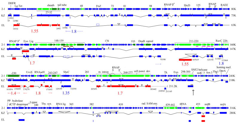
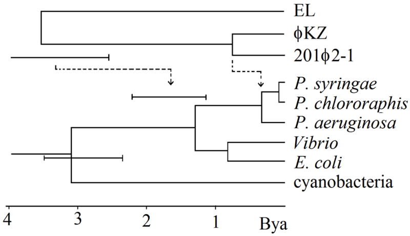
Pseudomonas chlororaphis phage 201varphi2-1 is a relative of Pseudomonas aeruginosa myovirus phiKZ. Phage 201 phi2-1 was examined by complete genomic sequencing (316,674 bp), by a comprehensive mass spectrometry survey of its virion proteins and by electron microscopy. Seventy-six proteins, of which at least 69 have homologues in phiKZ, were identified by mass spectrometry. Eight proteins, in addition to the major head, tail sheath and tail tube proteins, are abundant in the virion. Electron microscopy of 201 phi2-1 revealed a multitude of long, fine fibers apparently decorating the tail sheath protein. Among the less abundant virion proteins are three homologues to RNA polymerase beta or beta' subunits. Comparison between the genomes of 201 phi2-1 and phiKZ revealed substantial conservation of the genome plan, and a large region with an especially high rate of gene replacement. The phiKZ-like phages exhibited a two-fold higher rate of divergence than for T4-like phages or host genomes.


Figures

Figure 1
Figure 1
Virion proteins of 201φ2-1 separated by SDS-PAGE in a Bio-Rad Tris-HCl-buffered gradient gel (8 to 16% polyacrylamide). Proteins were visualized by staining with Coomassie Brilliant Blue. Molecular weights corresponding to the Precision Plus Protein Standard (Bio-Rad) are marked to the left of the sample lane. Identities of 201φ2-1 gene products assigned to bands are indicated to the right. In several cases, more than one protein was present in a gel band; these proteins are listed from highest to lowest relative abundance, left to right, based on spectrum counting (see Methods). Proteins migrating significantly faster than predicted from their molecular weights are marked with “*” or “” based on whether N- or C-terminal sequences, respectively, lacked MS sequence coverage.
Figure 2
Figure 2
Electron micrographs of 201φ2-1. Black short tailed arrows in all panels highlight several fine fibers projecting from the tail sheaths or heads of 201φ2-1 virions. (Top panel) Two particles of purified 201φ2-1 that were treated with DNAase immediately prior to being negatively stained with uranyl acetate. (Middle panel) Negatively stained 201φ2-1 without prior DNAase treatment. The arrow marked “a” indicates a possible strand of DNA. The inset image highlights a well stained baseplate from a virion. (Bottom panel) Cryo-EM image of 201φ2-1, without DNAase treatment. The white short tailed arrow highlights a particle (at the image edge) that has a contracted tail sheath and an empty head. The large white arrow highlights gasseous bubbles which formed in the head despite the use of low-dose techniques (see text). In the top and middle rows, large black arrows highlight particles that copurified with 201φ2-1 virions. In the bottom row, the large gray arrow highlights contamination on the cryo-EM specimen. All three scale bars = 100 nm.
Figure 3
Figure 3
Genome map of 201φ2-1 in comparison to φKZ and EL. Color coding in the 201φ2-1 track: green - gene product detected by MS (with abundant proteins in dark green); brown and magenta - two families of paralogues. Color coding in the KZ and EL tracks: blue - same orientation as the homologous 201φ2-1 gene, red - opposite orientation. Solid boxes are matching segments in the style of tblastx (see methods). An asterisk below an inderlined section of a φKZ match indicates protein sequence similarity within a newly predicted gene in φKZ (Supplementary Table 1). Hollow boxes indicate similarity detection requiring a profile method. The names of individual φKZ or EL genes found within the matching segments are in GenBank EU197055. The connectors between matching segments are inflected to indicate the amount of DNA inserted or deleted relative to 201φ2-1. Coordinates within the φKZ or EL genomes are given at the ends of chains of matches. The average 201φ2-1:EL/201φ2-1: φKZ divergence over segments delimited by inversion points is given in the same color as the chain of matches defining the segment and located below the EL track. The black dotted line for EL portrays the end of its genome relative to its rightmost major matching segment. Boxes not connected into chains map to uncorrelated positions in the different genomes. The vertical arrow indicates the beginning of the region discussed for its high rate of gene exchange in the text. Abbreviations are Lg. Ter (large subunit of terminase), RNAP (RNA polymerase), Exo. (exonuclease), Lig. (ligase), m. intron (mobile intron), Tk (thymidine kinase), Thy. syn. (thymidylate synthetase), rad. SAM enz. (radical SAM enzyme).
Figure 4
Figure 4
Phylogenetic tree relating the split times of the φKZ-like phages to the history of relevant bacterial species. The host tree was derived from rRNA sequence (see methods). The φKZ-like phage tree is shown either with an equal rate assumption (solid lines), or as adjusted for a two-fold greater divergence rate (dashed lines).

References

    1. Battistuzzi FU, Feijao A, Hedges SB. A genomic timescale of prokaryote evolution: insights into the origin of methaongenesis, phototrophy, and the colonization of land. BMC Evol Biol. 2004;4:44. - PMC - PubMed
    1. Bramucci MG, Keggins KM, Lovett PS. Bacteriophage PMB12 conversion of the sporulation defect in RNA polymerase mutants of Bacillus subtilis. J Virol. 1977;24:194–200. - PMC - PubMed
    1. Briers Y, Volckaert G, Cornelissen A, Lagaert S, Michiels CW, Hertveldt K, Lavigne R. Muralytic activity and modular structure or the endolysins of Pseudomonas aeruginosa bacteriophages φKZ and EL. Mol Microbiol. 2007;65:1334–1344. - PubMed
    1. Clark S, Losick R, Pero J. New RNA polymerase from Bacillus subtilis infected with phage PBS2. Nature. 1974;252:21–24. - PubMed
    1. Comeau A, Bertrand C, Letarov A, Tétart F, Krisch H. Modular architecture of the T4 phage superfamily: A conserved core genome and a plastic periphery. Virology. 2007;362:384–396. - PubMed

Publication types

Associated data