. 2008 Aug;22(8):2723-33.
doi: 10.1096/fj.08-106997. Epub 2008 May 12.

Diesel exhaust particles induce oxidative stress, proinflammatory signaling, and P-glycoprotein up-regulation at the blood-brain barrier

Affiliations

Diesel exhaust particles induce oxidative stress, proinflammatory signaling, and P-glycoprotein up-regulation at the blood-brain barrier

Anika M S Hartz et al. FASEB J. 2008 Aug.

Abstract

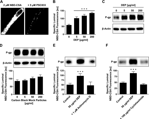
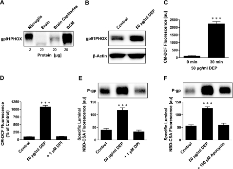
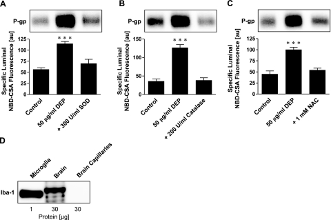
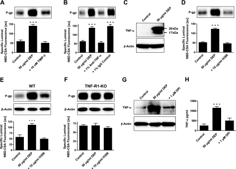
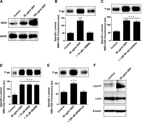
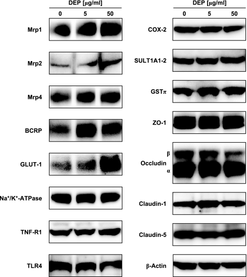
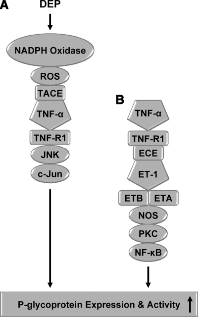
Here, we report that diesel exhaust particles (DEPs), a major constituent of urban air pollution, affect blood-brain barrier function at the tissue, cellular, and molecular levels. Isolated rat brain capillaries exposed to DEPs showed increased expression and transport activity of the key drug efflux transporter, P-glycoprotein (6 h EC(50) was approximately 5 microg/ml). Up-regulation of P-glycoprotein was abolished by blocking transcription or protein synthesis. Inhibition of NADPH oxidase or pretreatment of capillaries with radical scavengers ameliorated DEP-induced P-glycoprotein up-regulation, indicating a role for reactive oxygen species in signaling. DEP exposure also increased brain capillary tumor necrosis factor-alpha (TNF-alpha) levels. DEP-induced P-glycoprotein up-regulation was abolished when TNF-receptor 1 (TNF-R1) was blocked and was not evident in experiments with capillaries from TNF-R1 knockout mice. Inhibition of JNK, but not NF-kappaB, blocked DEP-induced P-glycoprotein up-regulation, indicating a role for AP-1 in the signaling pathway. Consistent with this, DEPs increased phosphorylation of c-jun. Together, our results show for the first time that a component of air pollution, DEPs, alters blood-brain barrier function through oxidative stress and proinflammatory cytokine production. These experiments disclose a novel blood-brain barrier signaling pathway, with clear implications for environmental toxicology, CNS pathology, and the pharmacotherapy of CNS disorders.


Figures

Figure 1.
Figure 1.
DEPs increase P-glycoprotein expression and transport activity in isolated rat brain capillaries. A) Left panel: representative image of an isolated brain capillary after 1 h of exposure to 2 μM NBD-CSA, showing steady-state NBD-CSA fluorescence. Note that NBD-CSA transport is concentrative from bath (no visible fluorescence) to endothelium (low fluorescence) to capillary lumen (high fluorescence). Right panel: the P-glycoprotein-specific inhibitor PSC833 blocks concentrative NBD-CSA transport from capillary endothelium to capillary lumen. B) DEPs increase specific luminal NBD-CSA fluorescence after 6 h in a concentration-dependent manner. C) Western blot analysis showing concentration-dependent P-glycoprotein up-regulation through DEPs in brain capillary plasma membranes. β-Actin was used as loading control. D) CB mock particles had no effect on P-glycoprotein expression or transport activity after 6 h. P-glycoprotein transport activity is shown as specific luminal NBD-CSA fluorescence. E) Inhibition of transcription with actinomycin D abolishes DEP-induced increase in P-glycoprotein expression (Western blot) and transport activity (specific luminal NBD-CSA fluorescence). F) Inhibition of protein synthesis with cycloheximide also abolishes DEP up-regulation of P-glycoprotein. For Specific luminal NBD-CSA fluorescence values are means ± se for 10 capillaries from a single preparation (pooled tissue from 10 rats); arbitrary units (au; scale 0–255). ***P < 0.001 vs. control.
Figure 2.
Figure 2.
NADPH oxidase and ROS are involved in DEP up-regulation of P-glycoprotein. A) Western blot showing expression of NADPH oxidase subunit gp91PHOX in microglia and brain capillary membranes. B) Exposing brain capillaries to DEPs for 6 h increased gp91PHOX expression in membranes. C) DEP exposure of brain capillaries for 30 min increased CM-DCF fluorescence, indicating increased ROS levels. ROS was measured using CM-H2DCFDA; values are means ± se (au; n=3) for capillaries from a single preparation (pooled tissue from 10 rats). D) NADPH oxidase inhibition with DPI abolished the DEP-induced increase of ROS (measured as CM-DCF fluorescence) in isolated brain capillaries. Data are percentages of control levels. E) Inhibition of NADPH oxidase with DPI blocked DEP up-regulation of P-glycoprotein expression (Western blot) and transport activity (specific luminal NBD-CSA fluorescence). F) Inhibition of NADPH oxidase with apocynin also blocked the DEP effect on P-glycoprotein. Specific luminal NBD-CSA fluorescence values are means ± se for 10 capillaries from a single preparation (pooled tissue from 10 rats); scale 0–255 au. ***P < 0.001.
Figure 3.
Figure 3.
ROS scavengers prevent P-glycoprotein up-regulation. A) The ROS scavenger SOD abolishes DEP up-regulation of P-glycoprotein expression (Western blot) and transport activity (specific luminal NBD-CSA fluorescence). B) Catalase, another ROS scavenger, also abolishes the effect of DEPs on P-glycoprotein. C) The antioxidant N-acetyl-l-cysteine (NAC) blocks DEP up-regulation of P-glycoprotein. D) Western blot showing expression of the microglia marker Iba-1. Iba-1 is expressed in microglia (positive control) and total brain, but not in brain capillaries. Specific luminal NBD-CSA fluorescence values are means ± se for 10 capillaries from a single preparation (pooled tissue from 10 rats); scale 0–255 au. ***P < 0.001.
Figure 4.
Figure 4.
DEP activation of NADPH oxidase is followed by TNF-α release and TNF-α signaling through TNF-R1 to up-regulate P-glycoprotein. A) TIMP-3, an inhibitor of TNF-α converting enzyme, blocks the DEP-mediated increase in P-glycoprotein expression (Western blot) and transport function (specific luminal NBD-CSA fluorescence). B) An antibody against TNF-α blocks the effect of DEPs on P-glycoprotein; an IgG control antibody has no effect. C) DEPs induce TNF-α in brain capillaries (26 kDa: TNF-α precursor protein, 17 kDa: TNF-α mature protein). D) Blocking TNF-R1 with H398 abolishes DEP up-regulation of P-glycoprotein. E) In isolated capillaries of wild-type mice, DEPs increase P-glycoprotein expression (Western blot) and transport activity (specific luminal NBD-CSA fluorescence). The specific TNF-R1 blocker, H398, blocks the DEP effect. F) In capillaries isolated from TNF-R1-deficient mice, DEPs had no effect on P-glycoprotein expression levels or transport activity. Note that different band intensities between controls in the Western blots of wild-type and TNF-R1 knockout mice result from different exposure times of the blotting membrane. G) Western blot showing that the NADPH oxidase inhibitor DPI blocks DEP up-regulation of P-glycoprotein. H) ELISA showing that blocking NADPH oxidase with DPI brings TNF-α levels back to control levels. Specific luminal NBD-CSA fluorescence values are means ± se for 10 capillaries from a single preparation (pooled tissue from 10 rats or 15 mice); scale 0–255 au. ***P < 0.001.
Figure 5.
Figure 5.
DEP up-regulation of P-glycoprotein involves NO synthase, JNK, and c-jun. A) Western blot showing that DEPs increase expression of iNOS, but not of bNOS. B) Inhibition of NOS with L-NMMA abolishes the DEP-mediated induction of P-glycoprotein expression and transport activity. C) An NF-κB activation inhibitor has no effect on DEP up-regulation of P-glycoprotein. D) SN50, an inhibitor of NF-κB nuclear translocation, and SN50M, the corresponding inactive control peptide, also have no effect on DEP up-regulation of P-glycoprotein. E) Inhibition of JNK with SP600125 blocks the DEP-mediated increase of P-glycoprotein expression (Western blot) and transport activity (specific luminal NBD-CSA fluorescence). F) Western blot showing that DEPs increase phosphorylated c-jun (c-jun-P) in capillaries exposed to DEPs for 6 h; c-jun expression was slightly decreased. Specific luminal NBD-CSA fluorescence values are means ± se for 10 capillaries from a single preparation (pooled tissue from 10 rats); scale 0–255 au. ***P < 0.001.
Figure 6.
Figure 6.
Effect of DEPs on the expression of selected proteins in isolated rat brain capillaries.
Figure 7.
Figure 7.
Comparison of DEP signaling (present study) and TNF-α signaling .

References

    1. Nel A. Atmosphere. Air pollution-related illness: effects of particles. Science. 2005;308:804–806. - PubMed
    1. U.N. Environment Programme, and W. H. O. Report Air pollution in the world’s megacities. Environment. 1994;36:2–13, 25–37.
    1. Singh P, DeMarini D M, Dick C A, Tabor D G, Ryan J V, Linak W P, Kobayashi T, Gilmour M I. Sample characterization of automobile and forklift diesel exhaust particles and comparative pulmonary toxicity in mice. Environ Health Perspect. 2004;112:820–825. - PMC - PubMed
    1. Draper W. Quantitation of nitro and dinitropolycyclic aromatic hydrocarbons in diesel exhaust particulate matter. Chemosphere. 1986;15:437–447.
    1. Schuetzle D. Sampling of vehicle emissions for chemical analysis and biological testing. Environ Health Perspect. 1983;47:65–80. - PMC - PubMed

Publication types

MeSH terms