. 2008 May 19;181(4):587-94.
doi: 10.1083/jcb.200803027. Epub 2008 May 12.

Molecular architecture of the kinetochore-microtubule attachment site is conserved between point and regional centromeres

Affiliations

Molecular architecture of the kinetochore-microtubule attachment site is conserved between point and regional centromeres

Ajit P Joglekar et al. J Cell Biol. .

Abstract

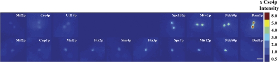
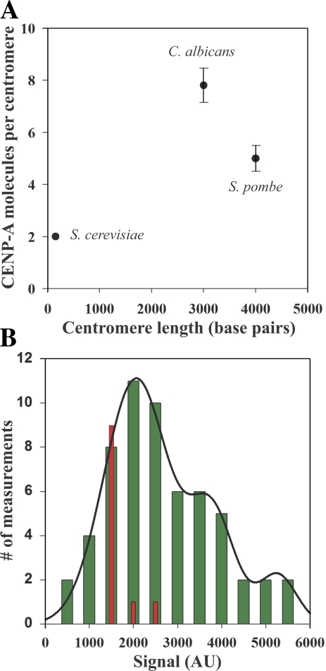
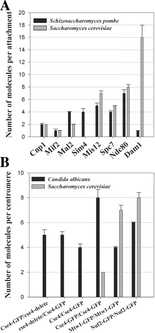
Point and regional centromeres specify a unique site on each chromosome for kinetochore assembly. The point centromere in budding yeast is a unique 150-bp DNA sequence, which supports a kinetochore with only one microtubule attachment. In contrast, regional centromeres are complex in architecture, can be up to 5 Mb in length, and typically support many kinetochore-microtubule attachments. We used quantitative fluorescence microscopy to count the number of core structural kinetochore protein complexes at the regional centromeres in fission yeast and Candida albicans. We find that the number of CENP-A nucleosomes at these centromeres reflects the number of kinetochore-microtubule attachments instead of their length. The numbers of kinetochore protein complexes per microtubule attachment are nearly identical to the numbers in a budding yeast kinetochore. These findings reveal that kinetochores with multiple microtubule attachments are mainly built by repeating a conserved structural subunit that is equivalent to a single microtubule attachment site.


Figures

Figure 1.
Figure 1.
Pseudo-colored images for a comparison of kinetochore protein fluorescence in metaphase budding yeast and G2/M fission yeast cells. Images were obtained under identical imaging and image acquisition conditions. Each of the two spots in a metaphase budding yeast cell contains 16 kinetochores and 16 microtubule attachment sites. In G2/M fission yeast, there are six kinetochores in the single spot with a mean of ∼3 microtubule attachment sites per kinetochore and, therefore, 18 attachment sites in total. It should be noted that microtubule attachments are established only after G2/M transition. The intensity bar is color coded to represent the ratio (indicated by the numbers on the right) with the ScCse4p-GFP intensity in the top panel. Bar, 1 μm.
Figure 2.
Figure 2.
The number of CENP-A molecules does not scale with the centromere DNA length. (A) The number of CENP-A molecules in the three fungi plotted as a function of centromere length. The number of CENP-A nucleosomes per centromere reflects the number of microtubule attachments per centromere. Error bars represent SD. (B) Signal measurement from clusters of one or more sister kinetochores in metaphase-arrested cells expressing SpNdc80p-GFP cells and a cold-sensitive nda3cs (β-tubulin) allele (green bars). A multipeak normal curve fit to the histogram (solid line) predicts peaks at 2,000, 3,800, and 5,600 intensity counts. Measurement of fluorescence signal from single lagging kinetochores in anaphase wild-type fission yeast expressing SpNuf2p-GFP yields a mean signal value of ∼1,000 counts (red bars). This confirms that the unit of minimum fluorescence signal in the nda3cs experiment corresponds to on pair of sister kinetochores. AU, arbitrary units.
Figure 3.
Figure 3.
Comparison of kinetochore protein numbers in the three fungi. (A) The number of core structural kinetochore proteins per microtubule attachment in fission yeast is very similar to the number of corresponding proteins at a budding yeast kinetochore. The Dam1–DASH complex, which is nonessential in fission yeast, is the sole exception (black bars, mean protein numbers in fission yeast; gray bars, mean numbers in budding yeast). (B) Although each C. albicans centromere contains approximately four CENP-A nucleosomes on average, the mean number of outer kinetochore proteins, such as Mtw1 and Nuf2, is sufficient to build only one microtubule attachment site (black bars, mean protein numbers in C. albicans; gray bars, mean numbers in budding yeast). Error bars represent SD from a minimum of two experiments.

References

    1. Black, B.E., L.E. Jansen, P.S. Maddox, D.R. Foltz, A.B. Desai, J.V. Shah, and D.W. Cleveland. 2007. Centromere identity maintained by nucleosomes assembled with histone H3 containing the CENP-A targeting domain. Mol. Cell. 25:309–322. - PubMed
    1. Bloom, K.S., E. Amaya, J. Carbon, L. Clarke, A. Hill, and E. Yeh. 1984. Chromatin conformation of yeast centromeres. J. Cell Biol. 99:1559–1568. - PMC - PubMed
    1. Blower, M.D., B.A. Sullivan, and G.H. Karpen. 2002. Conserved organization of centromeric chromatin in flies and humans. Dev. Cell. 2:319–330. - PMC - PubMed
    1. Bouck, D.C., and K.S. Bloom. 2005. The kinetochore protein Ndc10p is required for spindle stability and cytokinesis in yeast. Proc. Natl. Acad. Sci. USA. 102:5408–5413. - PMC - PubMed
    1. Bouck, D.C., and K. Bloom. 2007. Pericentric chromatin is an elastic component of the mitotic spindle. Curr. Biol. 17:741–748. - PMC - PubMed

Publication types