. 2008 Jun 9;205(6):1357-68.
doi: 10.1084/jem.20070950. Epub 2008 May 12.

Proteasomal degradation restricts the nuclear lifespan of AID

Affiliations

Proteasomal degradation restricts the nuclear lifespan of AID

Said Aoufouchi et al. J Exp Med. .

Abstract

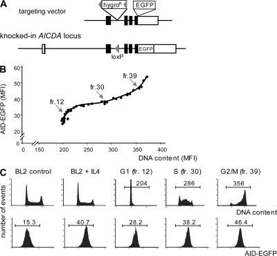
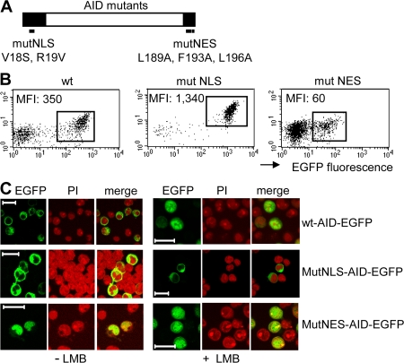
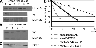
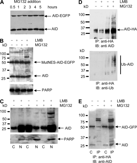
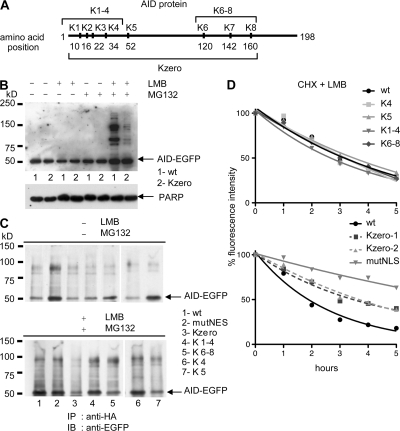
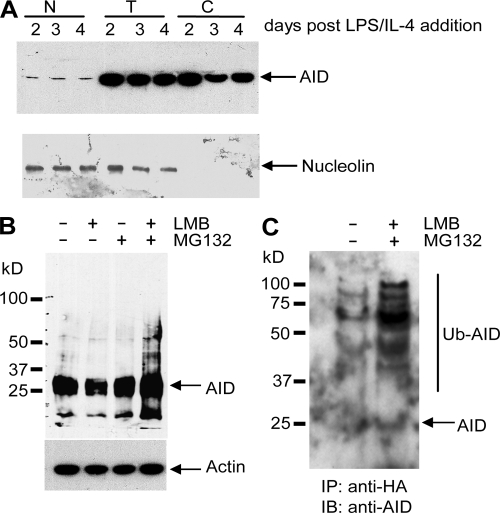
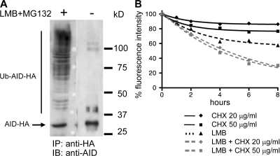
Activation-induced cytidine deaminase (AID) initiates all postrearrangement processes that diversify the immunoglobulin repertoire by specific deamination of cytidines at the immunoglobulin (Ig) locus. As uncontrolled expression of AID is potentially mutagenic, different types of regulation, particularly nucleocytoplasmic shuttling, restrict the likelihood of AID-deoxyribonucleic acid encounters. We studied additional mechanisms of regulation affecting the stability of the AID protein. No modulation of protein accumulation according to the cell cycle was observed in a Burkitt's lymphoma cell line. In contrast, the half-life of AID was markedly reduced in the nucleus, and this destabilization was accompanied by a polyubiquitination that was revealed in the presence of proteasome inhibitors. The same compartment-specific degradation was observed in activated mouse B cells, and also in a non-B cell line. No specific lysine residues could be linked to this degradation, so it remains unclear whether polyubiquitination proceeds through several alternatives sites or through the protein N terminus. The nuclear-restricted form of AID displayed enhanced mutagenicity at both Ig and non-Ig loci, most notably at TP53, suggesting that modulation of nuclear AID content through proteasomal degradation may represent another level of control of AID activity.


Figures

Figure 1.
Figure 1.
Variation of AID expression during the cell cycle. (A) EGFP KI at the AICDA locus in BL2 cells. The AID–EGFP KI construct includes the EGFP sequence inserted in-frame in exon 5 at the 3′ end of the AID coding region and a hygromycin resistance (hygroR) gene flanked by loxP sites. Configuration of the targeted AICDA locus is depicted after Cre-mediated excision of the hygroR gene. (B) Expression of AID-EGFP throughout the cell cycle. 48 h after IL-4 addition (10 ng/ml), AID-EGFP KI BL2 cells were fractionated according to their cell cycle status using counterflow elutriation. Collected fractions were stained with propidium iodide and analyzed for both DNA content and AID-EGFP MFI. Data for fractions 12, 30, and 39 are shown in C. (C) Cell cycle analysis and AID-EGFP expression level of the BL2 KI cell line with and without IL-4, and of representative elutriated fractions, corresponding to the different phases of cell cycle: G1 (fr.12), S (fr. 30), and G2/M (fr. 39).
Figure 2.
Figure 2.
Expression level of EGFP-tagged AID mutant proteins correlates with the specific cell compartment in which they reside. (A) Schematic representation of mutations affecting either the NLS (mutNLS, V18S, R19V, according to Shinkura et al. [13], with numbers indicating amino acid positions within the AID protein) or the NES (mutNES, L189A, F193A, and L196A), with both domains being represented as black boxes. (B) FACS analysis of the tetracycline-inducible clones, with values of the MFI of the EGFP-positive population. By comparison with endogenous AID expression levels in BL2 (corresponding to a MFI value of 15 in the KI clone [Fig. 1 C]), AID overexpression in EGFP-positive cells can be estimated at ∼4 for mutNES-AID, 24 for WT-AID, and 90 for mutNLS-AID constructs. (C) Confocal images of EGFP signals from inducible clones, with or without 2-h LMB treatment, are shown together with nuclear staining with propidium iodide (PI) of the same field. Clones with a lower percentage of inducible expression are shown, as indicated by the presence of EGFP-negative cells in the field, a shut-off of expression regularly observed in clones kept in culture in induced conditions. Bars, 20 μm.
Figure 3.
Figure 3.
The half-life of AID differs according to its subcellular localization. (A and B) BL2 clones expressing WT-AID-EGFP, MutNES-AID-EGFP, or MutNLS-AID-EGFP were metabolically labeled for 1 h with [35S]-labeled cysteine and methionine and chased for the indicated length of time. AID-EGFP was immunoprecipitated from cell lysates with anti-EGFP antibody (or anti-AID for the endogenous protein) and subjected to SDS-PAGE and autoradiography. (C) Immunoprecipitated MutNES-AID-EGFP was transferred into nitrocellulose and probed with a monoclonal HRP-conjugated anti-EGFP antibody to control for immunoprecipitation efficiency. (D) Densitometry analysis of autoradiographs of pulse-chase immunoprecipitated products, expressed as percentage of initial labeling. The data shown are the mean of the results from two experiments.
Figure 4.
Figure 4.
Ubiquitination of AID and AID-EGFP requires both nuclear localization and proteasome inhibition. (A) AID-EGFP knocked-in BL2 cells, expressing both AID and AID-EGFP under its endogenous promoter, were incubated with MG132 to inhibit proteasome activity. Cell lysates from the indicated time points were analyzed by immunoblotting using anti-AID monoclonal antibodies. (B) BL2 cells expressing both endogenous AID and MutNES-AID-EGFP were incubated for 5 h with LMB and/or MG132, as indicated. AID status was analyzed as described in A. Poly(ADP-ribose) polymerase (PARP; 116 kD) was used as loading control. (C) BL2 cells were incubated with LMB, MG132, or both, as indicated. For each condition, cytoplasmic (C) and nuclear (N) protein extracts were prepared as described in Materials and methods and analyzed by immunoblotting, with monoclonal anti-AID antibodies. Anti-PARP antibody was used as a nuclear protein control. (D) A BL2 cell line expressing WT-AID-HA was incubated with LMB or MG132, or both, for 5 h. After treatment, cell lysates were denatured and immunoprecipitated with agarose-conjugated anti-HA antibodies before SDS-PAGE separation and probing with anti-AID (top) or antiubiquitin antibodies (bottom). MW markers and migration positions of AID-HA are indicated. (E) AID-EGFP knocked-in BL2 cells were transfected with an HA-tagged ubiquitin-expressing vector and either incubated or not with LMB and MG132 for 5 h. Immunoprecipitation was performed 16 h after transfection, as described in D. Western blot analysis was performed using anti-AID antibodies. Lanes: C, extract before immunoprecipitation (1/10th of total extract); IP, immunoprecipitated proteins.
Figure 5.
Figure 5.
Lysine mutants of AID are polyubiquitinated in BL2. (A) Schematic presentation of the human AID protein sequence along with the positions of the lysine residues mutated in the various constructs. (B) BL2 clones expressing either WT-AID-EGFP (sample 1, WT), or a lysineless mutant of AID (Kzero-AID-EGFP; sample 2, Kzero) were incubated with the indicated inhibitors for 5 h. Protein extracts were analyzed by immunoblotting with anti-AID. PARP (116 kD) was used as loading control. (C) BL2 cell clones stably expressing either WT-AID-EGFP (WT), mutNES-AID-EGFP (mutNES), a lysineless mutant of AID (Kzero), or the four other AID-EGFP lysine mutants depicted in A were transiently transfected with an HA-tagged ubiquitin-expressing vector and either left untreated (top) or incubated with both LMB and MG132 for 5 h (bottom). Denatured lysates were immunoprecipitated with agarose-conjugated anti-HA antibodies and analyzed by Western blotting using anti-EGFP antibody. Migration position of AID-EGFP is indicated. (D) Stable BL2 clones expressing the indicated AID-EGFP lysine mutants, WT-AID-EGFP, or mutNLS-AID-EGFP were incubated with cycloheximide (CHX, 50 μg/ml) and LMB (10 ng/ml). The decay of the protein was followed by the decrease in fluorescence intensity. The percentage of initial fluorescence is plotted at various time points after addition of the drugs. Kzero1 and 2 represent two independent clones. (top) Clones transfected with AID constructs cloned in the pIRES puro expression vector; (bottom) clones transfected with tetracycline-inducible pBI expression vectors.
Figure 6.
Figure 6.
AID is ubiquitinated in the nucleus of activated mouse B splenocytes. (A) Purified B cells from mouse spleen were activated by incubation with LPS and IL-4. At the indicated days after activation, cells were collected and total (T), cytoplasmic (C), or nuclear (N) protein extracts were prepared as described in Materials and methods and analyzed by immunoblotting, with a monoclonal anti–mouse AID antibody. Antinucleolin antibody was used as a nuclear protein control. (B) At day 4, activated B cells were treated with LMB and/or MG132, as indicated. SDS-PAGE–fractionated extracts were probed with anti–mouse AID antibody. Actin was used as loading control. (C) At day 2 of IL-4/LPS activation, 2 × 10⁷ cells were transfected with Ub-HA–expressing vector using Amaxa nucleofection, as indicated in Materials and methods, and either left untreated or incubated for 4 h with both MG132 and LMB, 16 h after transfection. After treatment, cell lysates were denatured and immunoprecipitated with agarose-conjugated anti-HA antibodies before Western blot analysis with anti-AID monoclonal antibodies. MW markers and migration positions of endogenous AID are indicated.
Figure 7.
Figure 7.
Nuclear destabilization of AID is not restricted to B cells. (A) Human 293T cells transfected with WT-AID-HA–expressing vector were incubated for 6 h with both LMB and MG132, or left untreated, as indicated. Immunoprecipitation was performed using agarose-conjugated anti-HA antibodies, and SDS-PAGE–fractionated protein extracts were analyzed by Western blot with anti-AID antibodies. Migration position of AID-HA is indicated. (B) 293T cells stably transfected with a WT-AID-EGFP–expressing vector were incubated with cycloheximide (CHX; 20 or 50 μg/ml) and LMB, either separately or in combination, and the decay of the protein was followed by the decrease in fluorescence intensity. The percentage of initial fluorescence is plotted at various time points after addition of the inhibitors of protein synthesis and/or protein nuclear export.

References

    1. Jung, D., C. Giallourakis, R. Mostoslavsky, and F.W. Alt. 2006. Mechanism and control of V(D)J recombination at the immunoglobulin heavy chain locus. Annu. Rev. Immunol. 24:541–570. - PubMed
    1. Li, Z., C.J. Woo, M.D. Iglesias-Ussel, D. Ronai, and M.D. Scharff. 2004. The generation of antibody diversity through somatic hypermutation and class switch recombination. Genes Dev. 18:1–11. - PubMed
    1. Muramatsu, M., V.S. Sankaranand, S. Anant, M. Sugai, K. Kinoshita, N.O. Davidson, and T. Honjo. 1999. Specific expression of activation-induced cytidine deaminase (AID), a novel member of the RNA-editing deaminase family in germinal center B cells. J. Biol. Chem. 274:18470–18476. - PubMed
    1. Muramatsu, M., K. Kinoshita, S. Fagarasan, S. Yamada, Y. Shinkai, and T. Honjo. 2000. Class switch recombination and hypermutation require activation-induced cytidine deaminase (AID), a potential RNA editing enzyme. Cell. 102:553–563. - PubMed
    1. Revy, P., T. Muto, Y. Levy, F. Geissmann, A. Plebani, O. Sanal, N. Catalan, M. Forveille, R. Dufourcq-Labelouse, A. Gennery, et al. 2000. Activation-induced cytidine deaminase (AID) deficiency causes the autosomal recessive form of the Hyper-IgM syndrome (HIGM2). Cell. 102:565–575. - PubMed

Publication types