. 2008 Aug;76(8):3502-10.
doi: 10.1128/IAI.00226-08. Epub 2008 May 12.

MglA and Igl proteins contribute to the modulation of Francisella tularensis live vaccine strain-containing phagosomes in murine macrophages

Affiliations

MglA and Igl proteins contribute to the modulation of Francisella tularensis live vaccine strain-containing phagosomes in murine macrophages

Linda Bönquist et al. Infect Immun. 2008 Aug.

Abstract

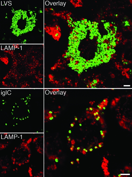
The Francisella tularensis live vaccine strain (LVS), in contrast to its iglC mutant, replicates in the cytoplasm of macrophages. We studied the outcome of infection of the murine macrophagelike cell line J774A.1 with LVS and with iglC, iglD, and mglA mutants, the latter of which is deficient in a global regulator. Compared to LVS, all of the mutants showed impaired intracellular replication up to 72 h, and the number of the mglA mutant bacteria even decreased. Colocalization with LAMP-1 was significantly increased for all mutants compared to LVS, indicating an impaired ability to escape into the cytoplasm. A lysosomal acidity-dependent dye accumulated in approximately 40% of the vacuoles containing mutant bacteria but not at all in vacuoles containing LVS. Preactivation of the macrophages with gamma interferon inhibited the intracellular growth of all strains and significantly increased acidification of phagosomes containing the mutants, but it only slightly increased the LAMP-1 colocalization. The intracellular replication and phagosomal escape of the iglC and iglD mutants were restored by complementation in trans. In conclusion, the IglC, IglD, and MglA proteins each directly or indirectly critically contribute to the virulence of F. tularensis LVS, including its intracellular replication, cytoplasmic escape, and inhibition of acidification of the phagosomes.


Figures

FIG. 1.
FIG. 1.
Western blot showing the levels of IglB, IglC, and IglD proteins in the igl and mglA mutants and in the complemented igl and mglA mutants. The presence of the three proteins in the LVS, ΔiglC, ΔiglD, and ΔmglA strains is shown in the blot on the left, and the presence in the complemented mutant strains is shown in the blot on the right. Each lane was loaded with a lysate prepared from 3 × 10⁷ bacteria.
FIG. 2.
FIG. 2.
Numbers of F. tularensis bacteria in J774 cells. J774 cells were infected for 1 h with the indicated F. tularensis strains at an MOI of 5 without (A) or with (B) IFN-γ and then incubated for the indicated periods. The numbers of bacteria at 0, 24, 48, and 72 h are expressed as the mean ± standard error of the mean log10 CFU for triplicate wells in one of the three experiments performed. The letter “a” indicates that the number of bacteria was significantly lower than the number of LVS bacteria at 24, 48, and 72 h (P < 0.05, as determined by a one-way ANOVA). The exception was iglD with IFN-γ at 72 h (P < 0.24). The comparisons were performed separately for IFN-γ-treated cultures. The letter “b” indicates that the log10 number of CFU of the ΔmglA mutant was significantly less (P < 0.001) than the values for the igl mutants at 48 and 72 h. The letter “c” indicates that IFN-γ treatment significantly inhibited the growth of each strain at 24, 48, and 72 h (P < 0.01).
FIG. 3.
FIG. 3.
Cytopathogenicity of F. tularensis strains. J774 cells were infected for 1 h with the indicated F. tularensis strains at an MOI of 5 in the absence or presence of IFN-γ. Cytopathogenicity was estimated by assaying LDH activity in culture supernatants and was expressed as a percentage of the level for noninfected lysed cells. The bars indicate the means and the error bars indicate the standard errors of the means for triplicate wells from one of three experiments. Asterisks indicate that the cytopathogenicity levels were significantly higher than those of uninfected cells, as determined by one-way ANOVA (*, P < 0.05; **, P < 0.01; ***, P < 0.001). Comparisons were performed separately for IFN-γ-treated cultures.
FIG. 4.
FIG. 4.
Colocalization of GFP-expressing F. tularensis strains and late endosomal markers. J774 cells were infected for 1 h with live or formalin-killed F. tularensis strains at an MOI of 30, or latex beads were added at an MOI of 10 and, after washing, incubated for 3 h. Fixed specimens were labeled for either (A) the late endosomal and lysosomal marker LAMP-1 or (B) the lysosomal marker CatD. In the representative confocal images, green indicates bacteria or latex particles and red indicates the endocytic marker. Confocal images were acquired with the Leica SP2 confocal microscope (Leica Microsystems, Bensheim, Germany) and were assembled using Adobe Photoshop CS2 (Adobe Systems, San Jose, CA). LB, latex beads. Bars = 4 μm.
FIG. 5.
FIG. 5.
Colocalization of GFP-expressing F. tularensis strains and LAMP-1. J774 cells were infected for 1 h with F. tularensis strains at an MOI of 5 and then incubated for 36 h. Fixed slides were labeled for the late endosomal and lysosomal marker LAMP-1. In the representative confocal images of cells infected with either LVS-GFP or ΔiglC-GFP, bacteria are indicated by green and LAMP-1 is indicated by red. Confocal images were acquired with the Leica SP2 confocal microscope and were assembled using Adobe Photoshop CS2. Bars = 4 μm.
FIG. 6.
FIG. 6.
Electron micrographs of J774 cells infected for 1 h with either F. tularensis LVS (B) or the ΔiglC mutant (A) and then incubated for 20 h. Electron micrographs were acquired with the JEOL 1200 EX-II electron microscope (JEOL Ltd., Tokyo, Japan) and were assembled using Adobe Photoshop CS2. Bars = 0.2 μm.

References

    1. Abd, H., T. Johansson, I. Golovliov, G. Sandström, and M. Forsman. 2003. Survival and growth of Francisella tularensis in Acanthamoeba castellanii. Appl. Environ Microbiol. 69600-606. - PMC - PubMed
    1. Anthony, L. S., P. J. Morrissey, and F. E. Nano. 1992. Growth inhibition of Francisella tularensis live vaccine strain by IFN-gamma-activated macrophages is mediated by reactive nitrogen intermediates derived from l-arginine metabolism. J. Immunol. 1481829-1834. - PubMed
    1. Balagopal, A., A. S. MacFarlane, N. Mohapatra, S. Soni, J. S. Gunn, and L. S. Schlesinger. 2006. Characterization of the receptor-ligand pathways important for entry and survival of Francisella tularensis in human macrophages. Infect. Immun. 745114-5125. - PMC - PubMed
    1. Brotcke, A., D. S. Weiss, C. C. Kim, P. Chain, S. Malfatti, E. Garcia, and D. M. Monack. 2006. Identification of MglA-regulated genes reveals novel virulence factors in Francisella tularensis. Infect. Immun. 746642-6655. - PMC - PubMed
    1. Brumell, J. H., and S. Grinstein. 2004. Salmonella redirects phagosomal maturation. Curr. Opin. Microbiol. 778-84. - PubMed

Publication types

MeSH terms

LinkOut - more resources