. 2008 Jun 7;53(11):2991-3006.
doi: 10.1088/0031-9155/53/11/016. Epub 2008 May 12.

Motion estimation for cardiac emission tomography by optical flow methods

Affiliations

Motion estimation for cardiac emission tomography by optical flow methods

D R Gilland et al. Phys Med Biol. .

Abstract

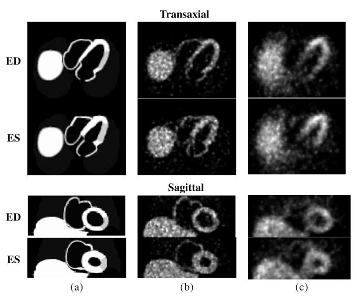
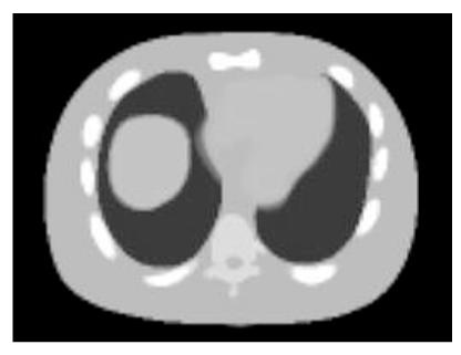
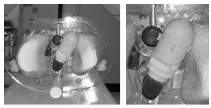
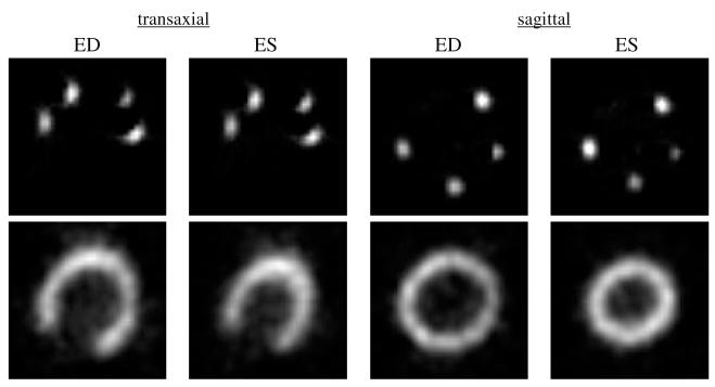
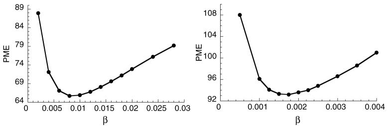
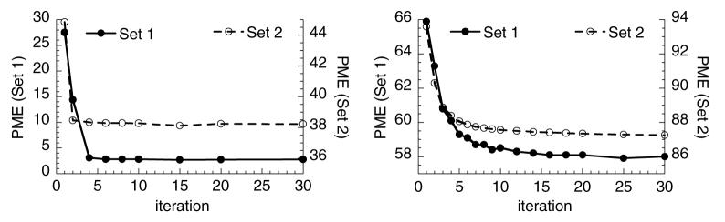
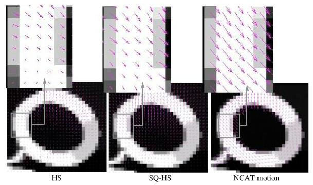
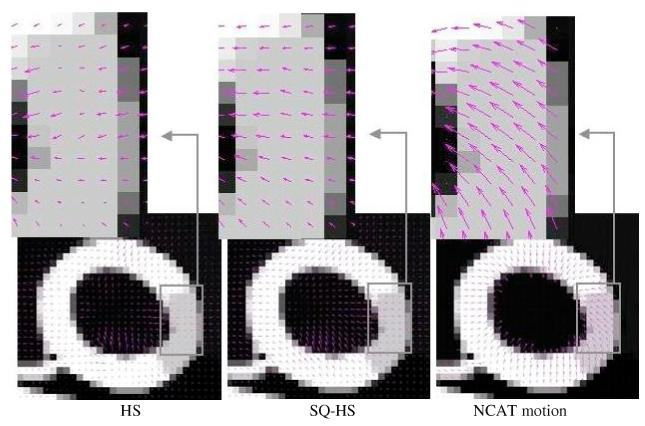
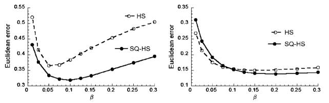
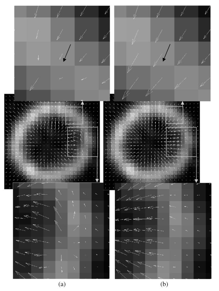
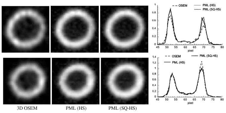
This paper describes a new method for estimating the 3D, non-rigid object motion in a time sequence of images. The method is a generalization of a standard optical flow algorithm that is incorporated into a successive quadratic approximation framework. The method was evaluated for gated cardiac emission tomography using images obtained from a mathematical, 4D phantom and a physical, dynamic phantom. The results showed that the proposed method offers improved motion estimation accuracy relative to the standard optical flow method. Convergence of the proposed algorithm was evidenced with a monotonically decreasing objective function value with iteration. Practical application of the motion estimation method in cardiac emission tomography includes quantitative myocardial motion estimation and 4D, motion-compensated image reconstruction.


Figures

Figure 1
Figure 1
Example 2D slice images of the 4D NCAT phantom in transaxial and sagittal slice orientations at end-diastole (ED) and end-systole (ES) frames. (a) Noise-free phantom, (b) Set 1 images (without attenuation, scatter and detector response effects) and (c) Set 2 images (with attenuation, scatter and detector response effects).
Figure 2
Figure 2
Transaxial slice of 4D NCAT attenuation distribution for Monte Carlo simulation.
Figure 3
Figure 3
Photograph of beating heart phantom showing myocardial markers. The outer shell of the phantom has been removed.
Figure 4
Figure 4
Reconstructed images of marker activity (top row) and myocardial activity (bottom row) for the physical, beating heart phantom.
Figure 5
Figure 5
One slice of the 4D region-of-interest (left) and the corresponding phantom image (right) used for the PME measurement.
Figure 6
Figure 6
Motion estimation error as a function of the penalty parameter β for the HS method. Left: Set 1 images and right: Set 2 images.
Figure 7
Figure 7
Motion estimation error as a function of the SQ-HS iteration number. Left: noise-free images and right: noisy images. In both plots, the left and right axes refer to Set 1 and Set 2 images, respectively.
Figure 8
Figure 8
Estimated motion vector fields from the Set 1 images superimposed on noise-free phantom images. The results are for a mid-systole frame interval (frame 1 to 2).
Figure 9
Figure 9
Estimated motion vector fields from the Set 2 images superimposed on noise-free images. The results are for a mid-systole frame interval (frame 1 to 2).
Figure 10
Figure 10
Objective function value versus iteration for the HS (left) and SQ-HS (right) methods.
Figure 11
Figure 11
Motion estimation error as a function of β for the physical phantom images. Left: frame 1-2 interval and right: averaged over all frame intervals.
Figure 12
Figure 12
Estimated motion vector fields superimposed on a reconstructed image of the physical phantom data for the frame 1-2 interval. (a) HS method and (b) SQ-HS method.
Figure 13
Figure 13
3D OSEM versus PML using the HS and SQ-HS motion estimates. Top row: frame 1, bottom row: frame 2. Profiles were taken along the middle, horizontal pixel row.

References

    1. Alvarez L, Weickert J, Sanchez J. Reliable estimation of dense optical flow fields with large displacements. Int. J. Comp. Vis. 2000;39:41–56.
    1. Black MJ, Anandan P. The robust estimation of multiple motions: parametric and piecewise-smooth flow fields. Comput. Vis. Image Understand. 1996;63:75–104.
    1. Brankov JG, Yang Y, Wernick MN. Spatiotemporal processing of gated cardiac SPECT images using deformable mesh modeling. Med. Phys. 2005;32:2839–49. - PubMed
    1. Cunningham GS, Hanson KM, Battle XL. Three-dimensional reconstructions from low-count SPECT data using deformable models. Opt. Exp. 1998;2:227–36. - PubMed
    1. Feng B, Bruyant PP, Pretorius PH, Beach RD, Gifford HC, Dey J, Gennert MA, King MA. Estimation of the rigid body motion from three dimensional images using a generalized center-of-mass points approach. IEEE Trans. Nucl. Sci. 2006;53:2712–8. - PMC - PubMed

Publication types

LinkOut - more resources