

The effect of voluntary fasting and dehydration on flicker-induced retinal vascular dilation in a healthy individual: a case report
- PMID: 18477404
- PMCID: PMC2408601
- DOI: 10.1186/1752-1947-2-153
The effect of voluntary fasting and dehydration on flicker-induced retinal vascular dilation in a healthy individual: a case report
Abstract
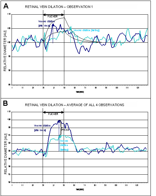
Introduction: Dynamic retinal vessel analysis represents a well-established method for the assessment of vascular reactivity during both normal conditions and after various provocations. We present a case where the subject showed abnormal retinal vessel reactivity after fasting voluntarily for 20 hours.
Case presentation: A healthy, 21-year-old man who fasted voluntarily for 20 hours exhibited abnormal retinal vascular reactivity (dilation and constriction) after flicker provocation as measured using the Dynamic Retinal Vessel Analyser (Imedos, Jena, Germany).
Conclusion: The abnormal vascular reactivity induced by fasting was significant; abnormal levels of important nutrients due to fasting and dehydration could play a role through altering the concentration of vasoactive substances such as nitric oxide. This hypothesis needs further investigation.
Figures


References
-
- Frederiksen CA, Jeppesen P, Knudsen ST, Poulsen PL, Mogensen CE, Bek T. The blood pressure-induced diameter response of retinal arterioles decreases with increasing diabetic maculopathy. Graefes Arch Clin Exp Ophthalmol. 2006 - PubMed
-
- Vilser W, Nagel E, Lanzl I. Retinal Vessel Analysis--new possibilities. Biomed Tech (Berl) 2002;47 Suppl 1 Pt 2:682–685. - PubMed
LinkOut - more resources
Full Text Sources
