. 2008 May;14(5):751-61.
doi: 10.1016/j.devcel.2008.02.015.

The C. elegans SYS-1 protein is a bona fide beta-catenin

Affiliations

The C. elegans SYS-1 protein is a bona fide beta-catenin

Jing Liu et al. Dev Cell. 2008 May.

Abstract

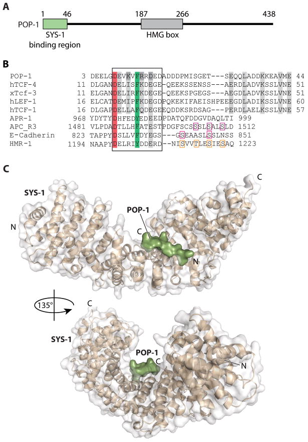
C. elegans SYS-1 has key functional characteristics of a canonical beta-catenin, but no significant sequence similarity. Here, we report the SYS-1 crystal structure, both on its own and in a complex with POP-1, the C. elegans TCF homolog. The two structures possess signature features of canonical beta-catenin and the beta-catenin/TCF complex that could not be predicted by sequence. Most importantly, SYS-1 bears 12 armadillo repeats and the SYS-1/POP-1 interface is anchored by a conserved salt-bridge, the "charged button." We also modeled structures for three other C. elegans beta-catenins to predict the molecular basis of their distinct binding properties. Finally, we generated a phylogenetic tree, using the region of highest structural similarity between SYS-1 and beta-catenin, and found that SYS-1 clusters robustly within the beta-catenin clade. We conclude that the SYS-1 protein belongs to the beta-catenin family and suggest that additional divergent beta-catenins await discovery.


Figures

Figure 1
Figure 1. SYS-1 contains a domain with 12 armadillo repeats
(A) SYS-1 domain structure predicted by FoldIndex. Folded and unfolded regions are colored in green and red, respectively. (B) Crystal structure of the SYS-1 armadillo repeat region. Each armadillo repeat (R1-R12) is given its own color. N, N-terminus; C, C-terminus. with each repeat colored. (C) Structure-based sequence alignment of the 12 repeats of C. elegans SYS-1. Repeat numbers and corresponding amino acids are shown on the left. The specific residues that form helices H1, H2, and H3 are boxed. Structural positions with strong preferences for a given amino acid or group of amino acids are shaded and listed on the line marked “Conserved” with the star symbols.
Figure 2
Figure 2. Comparison of armadillo repeat domains in SYS-1 and human β-catenin
(A) Superposition of residues 180–811 of SYS-1 (tan) and residues 134–664 of β-catenin (Protein Data Bank ID code 1G3J, slate) by Pymol. Both proteins are viewed from the side. See Supplementary Figure 1 for comparisons of individual armadillo repeats. (B) Top view of SYS-1 (left) and human β-catenin (right). This view is roughly related to that in (A) by a 90° rotation. The deeper groove of SYS-1 creates a hole when viewed from this angle. (C) Electrostatic surface representation of SYS-1 and β-catenin. Blue represents regions of positive potential and red represents regions of negative potential, at the 10 kT/e level. Both SYS-1 and canonical β-catenin carry a positively charged “button” area that is critical for POP-1/TCF binding. While POP-1 is largely neutral outside of this region, almost the entire β-catenin groove is positively charged. This figure was made with GRASP.
Figure 3
Figure 3. Structure of the SYS-1/POP-1 complex
(A) POP-1 protein has an N-terminal domain (green) that binds SYS-1 and a central domain (grey) that binds DNA. (B) Sequence alignment of β-catenin-binding domains that reside within a variety of β-catenin binding partners: C. elegans POP-1, human Tcf-4, Xenopus Tcf-3, human LEF-1, human Tcf-1, human APC (20 aa repeat 3; APC-R3), human E-cadherin, C. elegans HMR-1 (cadherin analogue) and C. elegans APR-1 (APC analogue). Framed by a rectangle is the conserved DxθθxΦx2–7E motif (θ and Φ are hydrophobic and aromatic residues, respectively), which is critical for the recognition of β-catenin repeats 5–9. The critical “charged button” and the conserved F/Y residues are colored in red and green, respectively. Other residues involved in β-catenin are also shaded. Critical phosphorylation sites observed in β-catenin/APC and β-catenin/E-cadherin crystal structures are framed in pink. Proposed HMR-1 phosphorylation sites are framed in beige. (C) Overall structure of the SYS-1/POP-1 complex. SYS-1(180–811) and POP-1(7–14) are in tan and green, respectively. Top view of SYS-1 corresponds roughly to that in figure 2A. Bottom view shows the shape of the groove that holds POP-1.
Figure 4
Figure 4. Crystal structure of SYS-1/POP-1 complex reveals the evolutionarily conserved “charged button”
(A) Representative region of the experimental electron density map of POP-1. The map was subjected to phase extension and density modification at 2.5 Å and contoured at 1 σ. SYS-1 is shown in electrostatic potential surface diagram. POP-1(3–18) is shown in green sticks. (B) Comparison of the “charged button” regions between the SYS-1/POP-1 and β-catenin/hTcf-4 complexes. The structures of these two complexes were superimposed. (C) Comparison of a critical hydrophobic interaction that is also evolutionarily conserved. (D) Association of SYS-1 with POP-1. Lane 1–4: GST(control), POP-1(1–200) and POP-1(1–45) tagged with GST were tested for its ability to bind wild-type SYS-1 (wt). Lanes 5–7: GST tagged POP-1(1–45) mutant (D8E) and SYS-1 containing point mutations in residues predicted to be important for interacting with POP-1 (K539A and A533L) were tested for interaction. GST-POP-1(1–45) was sufficient to interact with SYS-1 (lane 4), but POP-1(D8E), SYS-1(K539A) and SYS-1(A533L) all abolished the SYS-1/POP-1 interactions (Lane 5–7).
Figure 5
Figure 5. Structural explanation for the inability of WRM-1 to bind POP-1
(A) Secondary structure of R6-R8 of SYS-1 in sequence alignment with corresponding region of WRM-1. Helices are designated by repeat number (e.g. R6) and helix number (e.g. H1). (B) Superposition of the SYS-1/POP-1 complex together with selected amino acids from the WRM-1 model. The area around the charged button (K539 for SYS-1) is shown. The SYS-1(R6–R8) is shown in tan, WRM-1 corresponding region is in slate and the POP-1 N-terminal domain is in green. (C) Structure of the SYS-1/POP-1 complex. SYS-1 surface diagram is shown in tan, with residue A533 in green. (D) Surface diagram of the WRM-1 model. Residue L491 is shown in red. The fake POP-1 molecule as docked in panel (b) would collide with the side chain of L491.
Figure 6
Figure 6. SYS-1 clusters phylogenetically with other β-catenins
The unrooted neighbor joining tree was derived by PHYLIP using amino acid sequences for armadillo repeats 6–8 of: all four C. elegans β-catenins (SYS-1, WRM-1, HMP-2 and BAR-1), human β-catenin and two C. elegans importin-αhomologs, IMA-2 and IMA-3. Human importin-α2 was used as an outgroup. 1000 trees were generated and the bootstrap numbers on the branches indicate the percentage of trials the proteins partitioned into the two sets separated by that branch.

References

    1. Aberle H, Butz S, Stappert J, Weissig H, Kemler R, Hoschuetzky H. Assembly of the cadherin-catenin complex in vitro with recombinant proteins. J Cell Sci . 1994;107(Pt 12):3655–3663. - PubMed
    1. Chenna R, Sugawara H, Koike T, Lopez R, Gibson TJ, Higgins DG, Thompson JD. Multiple sequence alignment with the Clustal series of programs. Nucleic Acids Res. 2003;31:3497–3500. - PMC - PubMed
    1. Choi HJ, Huber AH, Weis WI. Thermodynamics of beta-catenin-ligand interactions: the roles of the N- and C-terminal tails in modulating binding affinity. J Biol Chem. 2006;281:1027–1038. - PubMed
    1. Costa M, Raich W, Agbunag C, Leung B, Hardin J, Priess JR. A putative catenin-cadherin system mediates morphogenesis of the Caenorhabditis elegans embryo. J Cell Biol. 1998;141:297–308. - PMC - PubMed
    1. Cox EA, Hardin J. Sticky worms: adhesion complexes in C. elegans. J Cell Sci. 2004;117:1885–1897. - PubMed

Publication types

MeSH terms