. 2008 May;14(5):762-74.
doi: 10.1016/j.devcel.2008.03.002.

Sensory signaling-dependent remodeling of olfactory cilia architecture in C. elegans

Affiliations

Sensory signaling-dependent remodeling of olfactory cilia architecture in C. elegans

Saikat Mukhopadhyay et al. Dev Cell. 2008 May.

Abstract

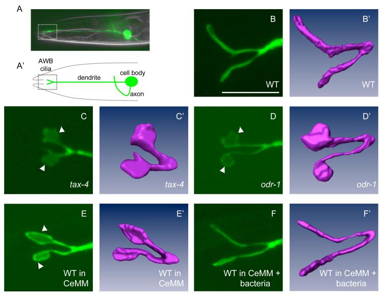
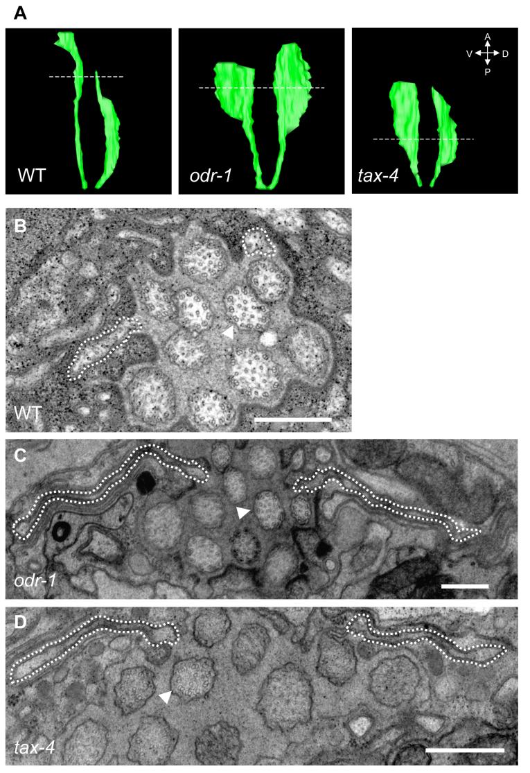
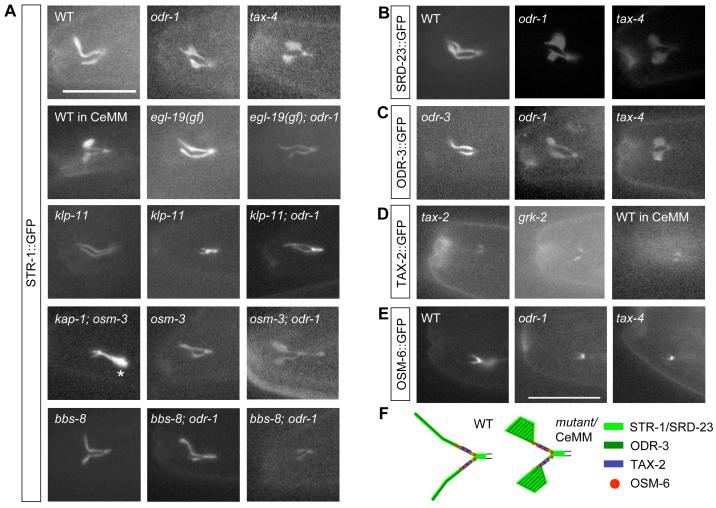
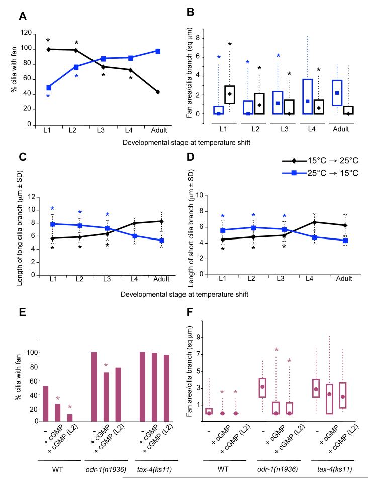
Nonmotile primary cilia are sensory organelles composed of a microtubular axoneme and a surrounding membrane sheath that houses signaling molecules. Optimal cellular function requires the precise regulation of axoneme assembly, membrane biogenesis, and signaling protein targeting and localization via as yet poorly understood mechanisms. Here, we show that sensory signaling is required to maintain the architecture of the specialized AWB olfactory neuron cilia in C. elegans. Decreased sensory signaling results in alteration of axoneme length and expansion of a membraneous structure, thereby altering the topological distribution of a subset of ciliary transmembrane signaling molecules. Signaling-regulated alteration of ciliary structures can be bypassed by modulation of intracellular cGMP or calcium levels and requires kinesin-II-driven intraflagellar transport (IFT), as well as BBS- and RAB8-related proteins. Our results suggest that compensatory mechanisms in response to altered levels of sensory activity modulate AWB cilia architecture, revealing remarkable plasticity in the regulation of cilia structure.


Figures

Figure 1
Figure 1
Sensory signaling modulates AWB cilia structure (A, A’) Location of the cell body, processes and cilia of an AWB olfactory neuron in the head of an adult animal. The AWB neuron is visualized via expression of str-1::gfp. Only one of the bilateral AWB neuron pair is visible in the shown lateral view. (B-F) The cilia of an AWB neuron visualized via str-1p::gfp transgene expression in wild-type (WT) adults grown under standard conditions (B, B’), tax-4(ks11) adults (C, C’), odr-1(n1936) adults (D, D’), wild-type adults grown in CeMM (E, E’), and wild-type adults grown in CeMM + bacteria (F, F’). All cilia images were acquired using confocal microscopy. (B’-F’) represent volumetric reconstructions from confocal projection series. Arrowheads indicate fans. Anterior is at left in all images. Scale - 7.5 μm for all confocal images.
Figure 2
Figure 2
Ultrastructure of AWB cilia in signaling mutants. (A) Three-dimensional reconstructions of the AWB cilia generated from serial EM sections obtained from wild-type, odr-1(n1936), and tax-4(ks11) mutants. The broken lines indicate the sections shown in (B-D). (B-D) A dashed line indicates the extent of the AWB cilia. Note that the axoneme structure of the channel cilia (one channel cilium indicated by arrowhead) is unaltered. The complete image sets of all acquired sections are shown in Movies S1-3. Scale - 500 nm.
Figure 3
Figure 3
Localization of ciliary proteins in AWB cilia. Shown are the localization patterns of GFP-tagged STR-1 (A), SRD-23 (B), ODR-3 (C), TAX-2 (D) and OSM-6 (E) in the cilia of an AWB neuron in the indicated growth conditions and genetic backgrounds. For klp-11 and bbs-8; odr-1 mutants, the cilia branch length phenotypes exhibited by a subset of these animals are indicated in a second panel in (A). Asterisk in (A) indicates accumulation at the ciliary base in kap-1; osm-3 double mutants. ODR-3::GFP and TAX-2::GFP rescue the corresponding mutant phenotypes, and their localization patterns in these mutant backgrounds are shown. OSM-6::GFP was previously shown to rescue the osm-6 phenotype in the AWB neurons (Mukhopadhyay et al., 2007). Alleles used were odr-1(n1936), tax-4(ks11), egl-19(n2368gf), klp-11(tm324), kap-1(ok676), bbs-8(nx77), osm-3(p802) and grk-2(gk268). WT - wild-type. Scale - 15 μm. (F) Summary of localization patterns of GFP-tagged proteins in AWB cilia.
Figure 4
Figure 4
TAX-2 and ODR-1 functions are required during later larval stages to modulate AWB cilia morphology. (A-D) tax-2(ks31) animals were raised at 15°C or 25°C and shifted to 25°C or 15°C respectively, at the indicated developmental stages. The percentage of cilia exhibiting fans (A), fan area per cilia branch (B), and lengths (±SD) of the long (C) and short (D) cilia branches were quantified. Asterisks mark values that are significantly different from those of animals grown throughout development at either 25°C (for animals shifted from 25°C to 15°C), or at 15°C (for animals shifted from 15°C to 25°C) at P<0.001. n = 30-100 for each time point. Box plots in (B) show the 25th, 50th (median, represented by symbol) and 75th percentiles, and the minimum and maximum values. (E-F) Modulation of cGMP levels alter AWB ciliary membrane area. Animals of the indicated genotypes were grown without or with 7 mM 8-Br-cGMP, and the percentage of cilia with fans (E) and fan areas (F) were quantified. 8-Br-cGMP was present either throughout development, or was added at the L2 larval stage. Shown are data from two or more independent experiments. Asterisks indicate values that are different from the corresponding strain grown in the absence of 8-Br-cGMP at P<0.001. n = 30-200 for each. Box plots in (F) show the 25th, 50th (median, represented by filled circles) and 75th percentiles, and the minimum and maximum values. All cilia measurements were performed in adult animals carrying stably integrated str-1p::gfp transgenes.

Comment in

References

    1. Alvarez VA, Sabatini BL. Anatomical and physiological plasticity of dendritic spines. Annu. Rev. Neurosci. 2007;30:79–97. - PubMed
    1. Ansley SJ, Badano JL, Blacque OE, Hill J, Hoskins BE, Leitch CC, Kim JC, Ross AJ, Eichers ER, Teslovich TM, et al. Basal body dysfunction is a likely cause of pleiotropic Bardet-Biedl syndrome. Nature. 2003;425:628–633. - PubMed
    1. Avery L. The genetics of feeding in Caenorhabditis elegans. Genetics. 1993;133:897–917. - PMC - PubMed
    1. Bargmann CI, Hartwieg E, Horvitz HR. Odorant-selective genes and neurons mediate olfaction in C. elegans. Cell. 1993;74:515–527. - PubMed
    1. Bargmann CI, Horvitz HR. Chemosensory neurons with overlapping functions direct chemotaxis to multiple chemicals in C. elegans. Neuron. 1991;7:729–742. - PubMed

Publication types

MeSH terms

LinkOut - more resources