. 2008 Jun 1;22(11):1513-21.
doi: 10.1101/gad.1661808. Epub 2008 May 13.

Dual KaiC-based oscillations constitute the circadian system of cyanobacteria

Affiliations

Dual KaiC-based oscillations constitute the circadian system of cyanobacteria

Yohko Kitayama et al. Genes Dev. .

Abstract

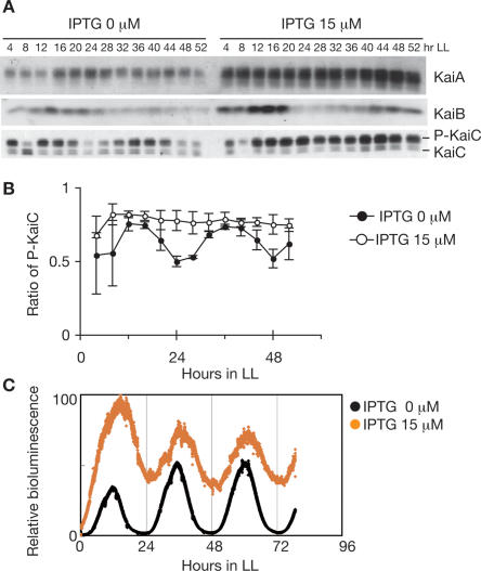
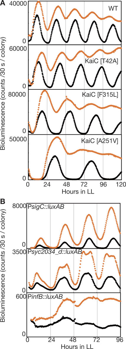
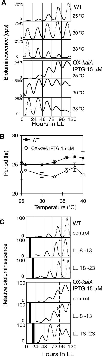
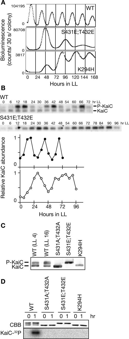
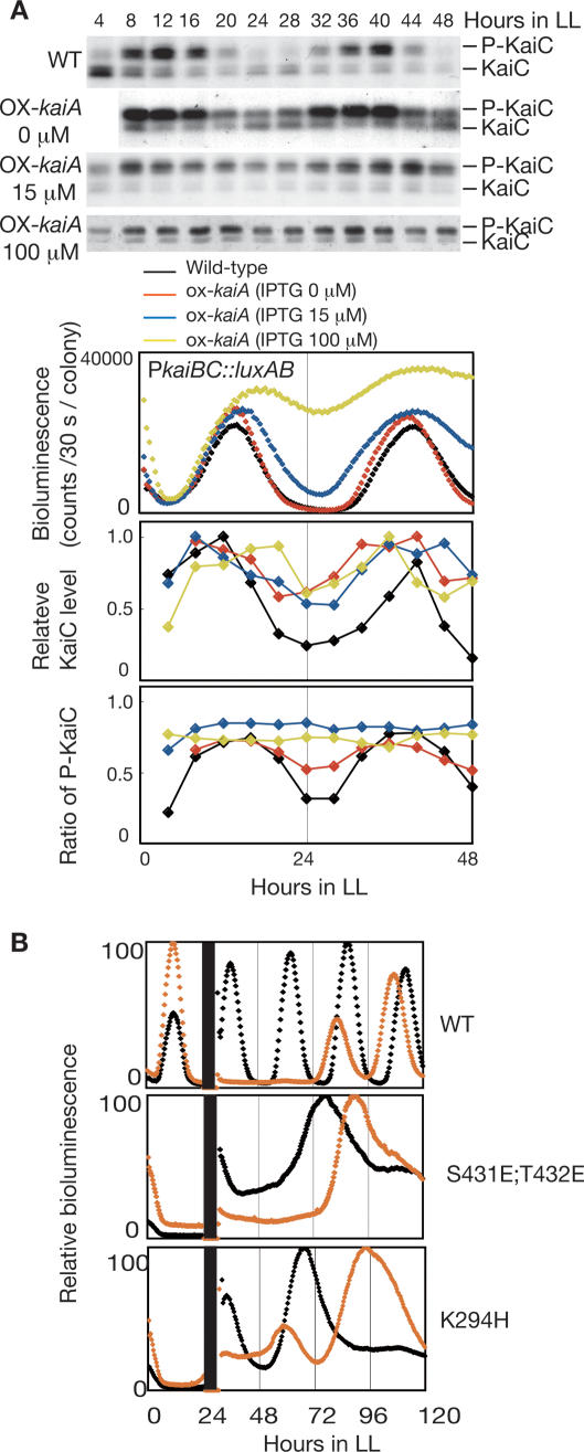
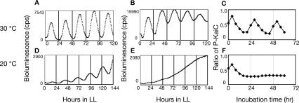
In the cyanobacterium Synechococcus elongatus PCC 7942, the KaiA, KaiB, and KaiC proteins are essential for the generation of circadian rhythms. Both in vivo and in vitro, phosphorylation of KaiC is regulated positively by KaiA and negatively by KaiB and shows circadian rhythmicity. The autonomous circadian cycling of KaiC phosphorylation is thought to be the basic pacemaker of the circadian clock and to control genome-wide gene expression in cyanobacteria. In this study, we found that temperature-compensated circadian oscillations of gene expression persisted even when KaiC was arrested in the phosphorylated state due to kaiA overexpression. Moreover, two phosphorylation mutants showed transcriptional oscillation with a long period. In kaiA-overexpressing and phosphorylation-deficient strains, KaiC oscillated and transient overexpression of phosphorylation-deficient kaiC reset the phase of the rhythm. These results suggest that transcription- and translation-based oscillations in KaiC abundance are also important for circadian rhythm generation in cyanobacteria. Furthermore, at low temperature, cyanobacteria can show circadian rhythms only when both the KaiC phosphorylation cycle and the transcription and translation cycle are intact. Our findings indicate that multiple coupled oscillatory systems based on the biochemical properties of KaiC are important to maintain robust and precise circadian rhythms in cyanobacteria.


Figures

Figure 1.
Figure 1.
Transcription–translation circadian oscillation in ox-kaiA. Cells were grown in a continuous culture system in continuous light (LL). After two 12-h light:12-h dark cycles, cells were returned to LL. Bioluminescence was measured continuously, and cells were collected at the indicated times. (A) Temporal profiles of Kai protein accumulation and KaiC phosphorylation in ox-kaiA cells harboring the PkaiBC∷luxAB reporter with or without 15 μM IPTG. Whole-cell extracts (2.5 μg for KaiA and KaiB, 1.5 μg for KaiC) were subjected to SDS-PAGE and Western blotting. In the bottom panel, the top band corresponds to phosphorylated KaiC (P-KaiC) and the bottom band corresponds to unphosphorylated KaiC (KaiC). (B) Quantification of KaiC phosphorylation in ox-kaiA cells with (open circles) or without (closed circles) 15 μM IPTG. The ratio of phosphorylated KaiC to total KaiC is plotted. The results are shown as means ± SEM (n = 4). (C) Bioluminescence rhythms of ox-kaiA cells under turbidostatic cultivation conditions with (orange circles) or without (black circles) 15 μM IPTG.
Figure 2.
Figure 2.
KaiC regulates global circadian transcription rhythms in ox-kaiA. (A) The bioluminescence rhythms of wild-type (WT) and three kaiC point mutants in kaiA-overexpressing cells. Bioluminescence was monitored on solid media with (orange diamonds) or without (black diamonds) 15 μM IPTG under LL. (B) The bioluminescence rhythms of three clones carrying promoter∷luxAB reporters isolated by promoter trapping from ox-kaiA cells. Bioluminescence was monitored on solid media with (orange diamonds) or without 15 μM IPTG (black diamonds) under LL. We confirmed that the upstream regions of sigC, syc2034_d (CYORF ID. Cyanobacteria Gene Annotation Database; http://cyano.genome.jp), or infB were fused to luxAB by sequencing the clones.
Figure 3.
Figure 3.
Oscillation without rhythmic KaiC phosphorylation has the characteristics of a circadian rhythm. (A) Temperature compensation of period length. Bioluminescence rhythms in the wild-type strain (top panel) and ox-kaiA strain induced with IPTG (bottom panel) at different temperatures. (B) The period of the bioluminescence rhythm was plotted against the culture temperature. Results are shown as means ± SEM (n = 3–6) for wild-type without IPTG (closed circles) and ox-kaiA induced with 15 μM IPTG (open circles). (C) Phase shifting of transcription–translation oscillation by a dark pulse. Cyanobacterial cells were inoculated onto agar plates and kept in LL for 1 d. After synchronization with a single 12-h dark period, the plates were returned to LL. Eight hours or 18 h after the return to LL, samples were placed in the dark for 5 h. After dark treatment, bioluminescence rhythms of wild-type (top panel) and ox-kaiA induced with IPTG (bottom panel) were monitored in LL. Maximum bioluminescence was standardized to 100. Black bars indicate the 5-h dark pulses.
Figure 4.
Figure 4.
Transcription–translation oscillation and phosphorylation. (A) Bioluminescence profiles of PkaiBC promoter activity in the wild-type strain (WT) and mutants in the phosphorylation sites [S431E;T432E] and in the Walker-A motif (K294H) are shown. Cells carrying the PkaiBC reporter cassette were grown on solid medium under LL. After a 12-h dark treatment, cells were returned to LL, and bioluminescence was measured. (B) Circadian profile of KaiC accumulation in wild-type and KaiC [S431E;T432E] mutant cells. Cells were grown in a continuous culture system in LL. After two 12-h light:12-h dark cycles, cells were returned to LL and collected at the indicated times. Whole-cell extracts (1.5 μg) were subjected to SDS-PAGE and Western blotting. The top band corresponds to phosphorylated KaiC (P-KaiC) and the bottom band corresponds to unphosphorylated KaiC (KaiC). Densitometric data for wild-type (closed circles) and KaiC [S431E;T432E] (open circles) are shown. Values at peak times were normalized to 1.0. (C) Phosphorylation state of KaiC protein mutated at the autophosphorylation sites ([S431A;T432A] and [S431E;T432E]) or the Walker-A motif (K294H). Proteins were extracted from wild-type (at LL4 and LL16) and mutant (at LL16) strains, and subjected to immunoblotting analysis to determine the phosphorylation state of KaiC. The top bands represent phosphorylated KaiC while the bottom bands represent unphosphorylated KaiC. (D) Autokinase activity of wild-type and KaiC mutant proteins. Wild-type and mutant KaiC were incubated with [γ-32P]ATP for 2 h in the presence of KaiA. The products were separated by SDS-PAGE followed by Coomassie Brilliant Blue (CBB)-staining (top panel) and autoradiography visualized using a BAS-2000 (bottom panel).
Figure 5.
Figure 5.
KaiC accumulation and phase resetting of gene expression rhythms. (A) Comparison of the changes in promoter activity, protein accumulation, and phosphorylation. Temporal profiles of bioluminescence, KaiC accumulation, and phosphorylation were examined in kaiA-overexpressing cells induced with 0, 15, or 100 μM IPTG, and wild-type cells. Whole-cell extracts (1.5 μg) were subjected to SDS-PAGE and Western blotting. The top band corresponds to phosphorylated KaiC (P-KaiC) and the bottom band corresponds to unphosphorylated KaiC (KaiC). Bioluminescence was measured under LL conditions. KaiC accumulation (middle) and KaiC phosphorylation (bottom) profiles were analyzed as in Figure 1A. Values at peak times were normalized to 1.0. The average accumulation of KaiC cannot be compared with each IPTG concentration because assays were performed by independent experiments with different exposure times. (B) Phase shifting after kaiC overexpression by IPTG induction. Bioluminescence rhythms of wild-type cells carrying a wild-type kaiC overexpression construct (top panel), KaiC [S431E;T432E] mutant cells carrying a kaiC [S431E;T432E] overexpression construct (middle panel), and KaiC [K294H] mutant cells carrying a kaiC [K294H] overexpression construct (bottom panel) are shown. Cells were treated with water (black line) or 1 mM IPTG (orange line) for 6 h at LL24.
Figure 6.
Figure 6.
Relationship between transcription–translation oscillation and phosphorylation oscillation. Bioluminescence profiles of wild-type cells (A,D), or ox-kaiA cells induced with 15 μM IPTG (B,E) under LL. Bioluminescence profiles were analyzed as in Figure 4A at 30°C (A,B) or 20°C (D,E). (C,F) KaiC phosphorylation profiles in vitro. Recombinant KaiC was incubated with KaiA and KaiB. Aliquots of reaction mixture were taken and subjected to SDS-PAGE and CBB staining. The ratio of phosphorylated KaiC to total KaiC is plotted. Assays were performed at 30°C (C) or 20°C (F).

Comment in

Similar articles

Cited by

References

    1. Arita K., Hashimoto H., Igari K., Akaboshi M., Kutusna S., Sato M., Shimizu T. Structural and biochemical characterization of a cyanobacterium circadian clock-modifier protein. J. Biol. Chem. 2007;282:1128–1135. - PubMed
    1. Bell-Pedersen D., Cassone V.M., Earnest D.J., Golden S.S., Hardin P.E., Thomas T.L., Zoran M.J. Circadian rhythms from multiple oscillators: Lessons from diverse organisms. Nat. Rev. Genet. 2005;6:544–556. - PMC - PubMed
    1. Clodong S., Duhring U., Kronk L., Wilde A., Axmann I., Herzel H., Kollmann M. Functioning and robustness of a bacterial circadian clock. Mol. Syst. Biol. 2007;3:90. doi: 10.1038/msb4100128. - DOI - PMC - PubMed
    1. Dunlap J.C., Loros J.J., DeCoursey P.J. Chronobiology: Biological timekeeping. Sinauer; Sunderland, MA: 2004.
    1. Emberly E., Wingreen N.S. Hourglass model for a protein-based circadian oscillator. Phys. Rev. Lett. 2006;96:038303. doi: 10.1103/PhysRevLett.96.038303. - DOI - PMC - PubMed

Publication types

Substances