. 2008 Jun 1;121(11):1852-60.
doi: 10.1242/jcs.012013. Epub 2008 May 13.

Identification of a novel N-terminal hydrophobic sequence that targets proteins to lipid droplets

Affiliations

Identification of a novel N-terminal hydrophobic sequence that targets proteins to lipid droplets

John K Zehmer et al. J Cell Sci. .

Abstract

AAM-B is a putative methyltransferase that is a resident protein of lipid droplets. We have identified an N-terminal 28 amino acid hydrophobic sequence that is necessary and sufficient for targeting the protein to droplets. This sequence will also insert AAM-B into the endoplasmic reticulum (ER). A similar hydrophobic sequence (1-23) in the cytochrome p450 2C9 cannot substitute for 1-28 and only inserts AAM-B into the ER, which indicates that hydrophobicity and ER anchoring are not sufficient to reach the droplet. We found that a similar N-terminal hydrophobic sequence in cytochrome b5 reductase 3 and ALDI could also heterologously target proteins to droplets. Targeting is not affected by changing a conserved proline residue that potentially facilitates the formation of a hairpin loop to leucine. By contrast, targeting is blocked when AAM-B amino acids 59-64 or 65-70, situated downstream of the hydrophobic sequence, are changed to alanines. AAM-B-GFP expressed in Saccharomyces cerevisiae is also faithfully targeted to lipid bodies, indicating that the targeting mechanism is evolutionarily conserved. In conclusion, a class of hydrophobic sequences exists that when placed at the N-terminus of a protein will cause it to accumulate in droplets and in the ER.


Figures

Fig. 1
Fig. 1
The AAM-B constructs used in this study and alignment of the N-terminal hydrophobic regions of AAM-B, ALDI, CYB5R3 and CYP2C9. (A) Wild-type AAM-B (WT) has four domains: an N-terminal hydrophobic region, a putative juxtamembrane domain, a putative methyltransferase domain and a C-terminal region that lacks any distinguishing features. Deletions made in the various regions as well as three chimeric proteins used in this study are shown. Each diagram represents a single construct, with the exception of AS1–7, which is a summary of seven constructs: the seven numbered boxes represent the regions mutated to alanines in the corresponding seven constructs. (B) Alignment of the first 23–28 amino acids of AAM-B, its close homolog ALDI, CYB5R3 and the p450 family member CYP2C9. Shaded boxes highlight hydrophobic residues (amino acid value>0 on the Kyte-Doolittle scale). Acidic residues are in red, basic residues in blue and prolines are highlighted yellow.
Fig. 2
Fig. 2
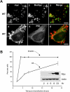
AAM-B is targeted to droplets. (A) HeLa cells were transfected with cDNA encoding C-terminally Myc-tagged AAM-B and grown for 15 hours in media containing 100 μM oleic acid to induce lipid droplets. The cells were fixed and processed for indirect immunofluorescence localization of Myc (1,4) and protein disulfide isomerase (5) or stained for lipids with Bodipy 493/503 (2). Most cells had Myc staining (1) restricted to the periphery of neutral lipid-positive (2) droplets, as shown in the merge (3). Other cells displayed Myc staining (4) around lipid droplets and, in addition, a reticular pattern distributed throughout the cell that co-localized with α-PDI IgG (5), as shown in the merge (6). (B) HeLa cells grown on coverslips in the absence of oleate were transfected with cDNAs encoding wild-type, Myc-tagged AAM-B. At various times post-transfection, the cells on the coverslip were fixed and processed for immunofluorescence detection of Myc. The cells remaining in the dish were collected, lysed and processed for immunoblotting to detect Myc (inset). To quantify the distribution of AAM-B, images of stained cells were systematically acquired until 50 cells had been photographed for each time point. The cells were then scored for the presence of either lipid droplet alone, or droplet plus reticular ER staining patterns. We did not detect any expressing cells until after a 2 hour incubation. Each point is the average ±s.e.m. of three separate experiments. Scale bars: 5 μm.
Fig. 3
Fig. 3
Deletion of amino acids 6–28 interrupts AAM-B targeting to droplets. The indicated AAM-B mutants were expressed in HeLa cells and processed for immunofluorescence. (A–C) Deletion of amino acids 2–18 (N2) did not disrupt the targeting of AAM-B (A) to Bodipy 493/503-positive (B) lipid droplets (C). (D–F) AAM-B lacking amino acids 6–28 (N3) had a cytosolic distribution (D). Brightly staining regions (arrows) appear to be insoluble aggregates that do not overlap with Bodipy 493/503-stained (E) lipid droplets (F). (G–I) AAM-B with amino acids 1–23 of CYP2C9 substituted for amino acids 1–28 (δN) had a reticular pattern (G) that was not even weakly associated with Bodipy-positive (H) droplets (I). Scale bars: 5 μm.
Fig. 4
Fig. 4
Amino acids 1–28 are sufficient to target proteins to droplets. AAM-B mutants were expressed in HeLa cells and processed for immunofluorescence. (A–C) Deletion of amino acids 29–61 in the juxtamembrane region (A) disrupted targeting to Bodipy 493/593-positive (B) droplets (C). (D–F) GFP was fused to the C-terminal end of AAM-B amino acids 1–28 and expressed in HeLa cells. The chimera (D) was found in an ER pattern with some protein localized to ADRP-marked (E) droplets (F). (G–I) AAM-B amino acids 1–38 fused to GFP (G) co-localized with ADRP (H) on droplets (I). (J–L) Deletion of amino acids 29–38 from full-length AAM-B (J) did not influence targeting to Bodipy-positive (K) droplets (L). Scale bars: 5 μm.
Fig. 5
Fig. 5
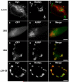
N-terminal hydrophobic sequences from other droplet proteins are sufficient to target droplets. (A–C) Amino acids 1–28 from AAM-B fused to Rab5 S34N (A) target Bodipy-positive (B) droplets (C). (D–F) Amino acids 1–27 of mouse ALDI fused to Rab5 S34N were sufficient to target the protein (D) to Bodipy-positive (E) droplets (F). (G–I) Protein comprising amino acids 1–28 of CYB5R3 fused to Rab5 S34N (G) was also found surrounding Bodipy-positive (H) droplets (I). Scale bars: 5 μm.
Fig. 6
Fig. 6
AAM-B targets to yeast lipid droplets. Cells were co-transformed with AAM-B-GFP and Erg6-DsRed and grown on selective plates. AAM-B-GFP (A,E) co-expressed with Erg6-DsRed (B,F) co-localized (C,G) on S. cerevisiae lipid bodies (shown under light microscopy in D,H). Scale bars: 5 μm.
Fig. 7
Fig. 7
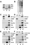
Cell fractionation shows that AAM-B is targeted to droplets. (A) Protease-protection assay shows that AAM-B is exposed to the cytosol. HeLa cells were transfected with GFP-AAM-B-Myc and equal aliquots of the total membranes were incubated in the absence (lane 1) or presence (lanes 2 and 3) of trypsin. Lane 3 contained a protease inhibitor. An immunoblot for calnexin (Cnx) shows that the protein is reduced by ~10 kDa, which corresponds to the size of the cytosolic part of the protein. GFP and Myc are digested in the presence of trypsin, demonstrating that they are cytosolically oriented. (B–F) COS7 cells were transfected with cDNAs encoding Myc-tagged AAM-B constructs and cell fractions prepared. Equal volumes of the droplet, cytosol and membrane fractions were separated by SDS-PAGE. (B) A representative gel was stained with Coomassie Blue to determine the relative protein load for the immunoblots in C–F. (C–F) Each preparation was immunoblotted with α-Myc, α-ADRP, α-Bip, α-Sec61β, α-PDI and α-actin IgG. (C) The wild-type protein was found in both droplet and membrane fractions. (D) The δN mutant was found solely in the membrane fraction. (E) The C6 mutant was found in the droplet fraction. (F) The ΔJxm mutant was found in the membrane fraction.
Fig. 8
Fig. 8
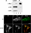
AAM-B on droplets can interact with itself. (A) Co-immunoprecipitation of Myc-tagged and HA-tagged AAM-B co-expressed in HeLa cells. HeLa cells were co-transfected with cDNA encoding either HA-tagged AAM-B, Myc-tagged AAM-B (lanes 2 and 4) or empty vector (lanes 1 and 3). The cells were solubilized with detergent and processed to immunoprecipitate AAM-B-Myc with α-Myc IgG. Samples of the cleared cell lysates (lanes 1 and 2) and the immunoprecipitates (lanes 3 and 4) were separated by SDS-PAGE and immunoblotted with α-HA IgG, α-Myc IgG or α-p63 IgG. (B–G) Wild-type AAM-B can recruit truncated AAM-B from the cytoplasm to droplets. HeLa cells were co-transfected with a cDNA encoding Myc-tagged N3 and an empty vector (B–D). Cells were grown for 15 hours, fixed and processed for immunofluorescence detection of HA (B) and Myc (C). HeLa cells were co-transfected with a cDNA encoding Myc-tagged N3 and HA-tagged wild-type AAM-B (E–G). Cells were grown for 15 hours, fixed and processed for immunofluorescence detection of HA (E) and Myc (F). Scale bars: 5 μm.

References

    1. Abell BM, Holbrook LA, Abenes M, Murphy DJ, Hills MJ, Moloney MM. Role of the proline knot motif in oleosin endoplasmic reticulum topology and oil body targeting. Plant Cell. 1997;9:1481–1493. - PMC - PubMed
    1. Bartz R, Zehmer JK, Zhu M, Chen Y, Serrero G, Zhao Y, Liu P. Dynamic activity of lipid droplets: protein phosphorylation and Gtp-mediated protein translocation. J. Proteome Res. 2007;6:3256–3265. - PubMed
    1. Binns D, Januszewski T, Chen Y, Hill J, Markin VS, Zhao Y, Gilpin C, Chapman KD, Anderson RG, Goodman JM. An intimate collaboration between peroxisomes and lipid bodies. J. Cell Biol. 2006;173:719–731. - PMC - PubMed
    1. Blanchette-Mackie EJ, Dwyer NK, Barber T, Coxey RA, Takeda T, Rondinone CM, Theodorakis JL, Greenberg AS, Londos C. Perilipin is located on the surface layer of intracellular lipid droplets in adipocytes. J. Lipid Res. 1995;36:1211–1226. - PubMed
    1. Bracey MH, Cravatt BF, Stevens RC. Structural commonalities among integral membrane enzymes. FEBS Lett. 2004;567:159–165. - PubMed

Publication types

MeSH terms