

DNA conformations and their sequence preferences
- PMID: 18477633
- PMCID: PMC2441783
- DOI: 10.1093/nar/gkn260
DNA conformations and their sequence preferences
Abstract
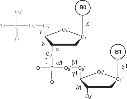
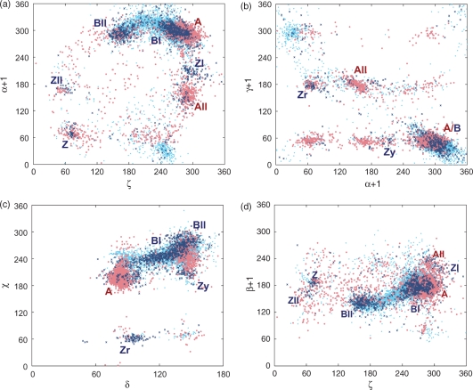
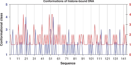
The geometry of the phosphodiester backbone was analyzed for 7739 dinucleotides from 447 selected crystal structures of naked and complexed DNA. Ten torsion angles of a near-dinucleotide unit have been studied by combining Fourier averaging and clustering. Besides the known variants of the A-, B- and Z-DNA forms, we have also identified combined A + B backbone-deformed conformers, e.g. with alpha/gamma switches, and a few conformers with a syn orientation of bases occurring e.g. in G-quadruplex structures. A plethora of A- and B-like conformers show a close relationship between the A- and B-form double helices. A comparison of the populations of the conformers occurring in naked and complexed DNA has revealed a significant broadening of the DNA conformational space in the complexes, but the conformers still remain within the limits defined by the A- and B- forms. Possible sequence preferences, important for sequence-dependent recognition, have been assessed for the main A and B conformers by means of statistical goodness-of-fit tests. The structural properties of the backbone in quadruplexes, junctions and histone-core particles are discussed in further detail.
Figures
References
-
- Schultz SC, Shields GC, Steitz TA. Crystal structure of a CAP-DNA complex: the DNA is bent by 90°. Science. 1991;253:1001–1007. - PubMed
-
- Luger K, Mader AW, Richmond RK, Sargent DF, Richmond TJ. Crystal structure of the nucleosome core particle at 2.8 A resolution. Nature. 1997;389:251–260. - PubMed
-
- Matthews BW. No code for recognition. Nature. 1988;335:294–295. - PubMed
-
- Pabo CO, Nekludova L. Geometric analysis and comparison of protein-DNA interfaces: why is there no simple code for recognition? J. Mol. Biol. 2000;301:597–624. - PubMed
Publication types
MeSH terms
Substances
LinkOut - more resources
Full Text Sources
Miscellaneous
