. 2008 May 27;105(21):7606-11.
doi: 10.1073/pnas.0802197105. Epub 2008 May 13.

Atomic interactions of neonicotinoid agonists with AChBP: molecular recognition of the distinctive electronegative pharmacophore

Affiliations

Atomic interactions of neonicotinoid agonists with AChBP: molecular recognition of the distinctive electronegative pharmacophore

Todd T Talley et al. Proc Natl Acad Sci U S A. .

Abstract

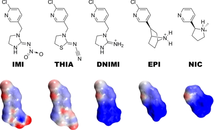
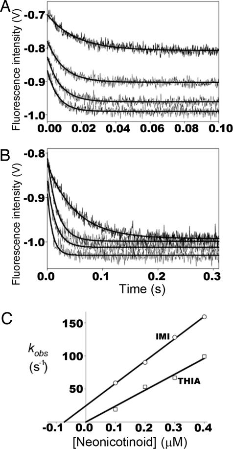
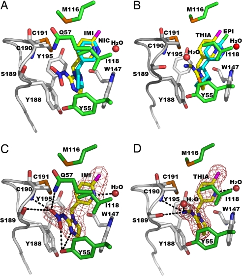
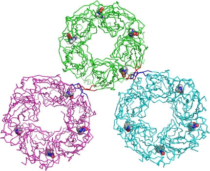
Acetylcholine-binding proteins (AChBPs) from mollusks are suitable structural and functional surrogates of the nicotinic acetylcholine receptors when combined with transmembrane spans of the nicotinic receptor. These proteins assemble as a pentamer with identical ACh binding sites at the subunit interfaces and show ligand specificities resembling those of the nicotinic receptor for agonists and antagonists. A subset of ligands, termed the neonicotinoids, exhibit specificity for insect nicotinic receptors and selective toxicity as insecticides. AChBPs are of neither mammalian nor insect origin and exhibit a distinctive pattern of selectivity for the neonicotinoid ligands. We define here the binding orientation and determinants of differential molecular recognition for the neonicotinoids and classical nicotinoids by estimates of kinetic and equilibrium binding parameters and crystallographic analysis. Neonicotinoid complex formation is rapid and accompanied by quenching of the AChBP tryptophan fluorescence. Comparisons of the neonicotinoids imidacloprid and thiacloprid in the binding site from Aplysia californica AChBP at 2.48 and 1.94 A in resolution reveal a single conformation of the bound ligands with four of the five sites occupied in the pentameric crystal structure. The neonicotinoid electronegative pharmacophore is nestled in an inverted direction compared with the nicotinoid cationic functionality at the subunit interfacial binding pocket. Characteristic of several agonists, loop C largely envelops the ligand, positioning aromatic side chains to interact optimally with conjugated and hydrophobic regions of the neonicotinoid. This template defines the association of interacting amino acids and their energetic contributions to the distinctive interactions of neonicotinoids.


Conflict of interest statement

The authors declare no conflict of interest.

Figures

Fig. 1.
Fig. 1.
Chemical structures of neonicotinoids imidacloprid (IMI) and thiacloprid (THIA) with an electronegative nitro or cyano pharmacophore compared with cationic nicotinic agonists desnitroimidacloprid (DNIMI), epibatidine (EPI), and nicotine (NIC). Below each structure is shown its charge distribution with the electron clouds in red for negative and blue for positive. Electrostatic potentials were calculated and images rendered by using Accelrys DS Visualizer v1.7.
Fig. 2.
Fig. 2.
Kinetic studies of ligand association with AChBP. Stopped-flow traces of tryptophan fluorescence quenching on neonicotinoid binding to Aplysia AChBP. Binding of 0.1, 0.2, 0.3, and 0.4 μM (ordered top to bottom) IMI (A) and THIA (B) to 4 nM AChBP pentamer. (C) First-order rate constants of fluorescence quenching (kobs) determined from A and B plotted against neonicotinoid concentration. Intercepts of lines on the ordinate represent first-order dissociation rate constants (koff), intercepts on the abscissa represent Kd, and line slopes represent second-order association rate constants (kon). For THIA only, kon could be determined from the association kinetics, whereas for IMI all three constants (kon, koff, and Kd) were estimated. For both IMI and THIA the first-order dissociation rate constants were measured in separate experiments by the enhancement of fluorescence after addition of excess gallamine or strychnine.
Fig. 3.
Fig. 3.
Bound agonists shown as overlapping or individual structures. (A) AChBP-IMI (yellow) and AChBP-NIC (PDB ID code 1UW6) (11) (cyan). (B) AChBP-THIA (yellow) and AChBP-EPI (PDB ID code 2BYQ) (4) (cyan). IMI (C) and THIA (D) electron density omit maps with coefficients, FoFc >3σ, showing hydrogen bonds as dashed lines. Principal face residues with distances <3.6 Å from the ligands and loop C are shown in gray. Complementary face residues are in given green, S atoms and Cys–Cys bond in orange, and Cl in magenta.
Fig. 4.
Fig. 4.
Overlap of loop C and the bound ligands in AChBP, AChBP-IMI, AChBP-THIA, and AChBP-EPI (4). (A) Subunit A with closed loop C conformation for the ligand-bound AChBP-IMI (red), AChBP-THIA (green), and AChBP-EPI (yellow). Loop C has an open conformation in the native AChBP (blue) structure. (B) Subunit E in which only AChBP-EPI has a bound ligand shows a closed loop C conformation (yellow), whereas the apo-AChBP, AChBP-IMI, and AChBP-THIA sites which are unoccupied show an open loop C conformation (see also Fig. 5).
Fig. 5.
Fig. 5.
Interactions of an AChBP-IMI molecule (green) with the two symmetry-related molecules that form intermolecular contacts <3.6 Å (shown in cyan and magenta). A closed loop C (red) of subunit A (green) forms a β-sheet with an open loop C (blue) of a symmetry-related molecule (cyan) of subunit E. A similar packing interaction is seen between an open loop C (red) of subunit E to a closed loop C (blue) of another symmetry-related molecule (magenta) of subunit A. The bound IMI molecules are shown as CPK models.

References

    1. Changeux JP, Edelstein SJ. Nicotinic Acetylcholine Receptors: From Molecular Biology to Cognition. New York: Odile Jacob/Johns Hopkins Univ Press; 2005.
    1. Karlin A. Emerging structure of the nicotinic acetylcholine receptors. Nat Rev Neurosci. 2002;3:102–114. - PubMed
    1. Talley TT, et al. Spectroscopic analysis of benzylidene anabaseine complexes with acetylcholine binding proteins as models for ligand-nicotinic receptor interactions. Biochemistry. 2006;45:8894–8902. - PMC - PubMed
    1. Hansen SB, et al. Structures of Aplysia AChBP complexes with nicotinic agonists and antagonists reveal distinctive binding interfaces and conformations. EMBO J. 2005;24:3635–3646. - PMC - PubMed
    1. Gotti C, Clementi F. Neuronal nicotinic receptors: From structure to pathology. Prog Neurobiol. 2004;74:363–396. - PubMed

Publication types

MeSH terms

LinkOut - more resources