. 2008 May 14;3(5):e2162.
doi: 10.1371/journal.pone.0002162.

Expression of aquaporin 5 (AQP5) promotes tumor invasion in human non small cell lung cancer

Affiliations

Expression of aquaporin 5 (AQP5) promotes tumor invasion in human non small cell lung cancer

Young Kwang Chae et al. PLoS One. .

Erratum in

Abstract

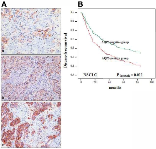
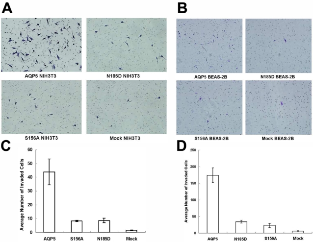
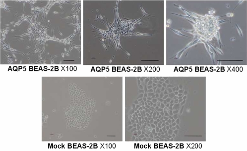
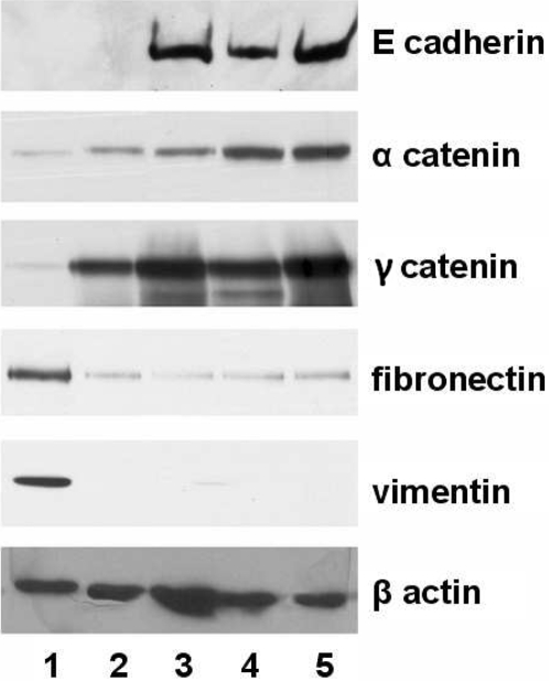
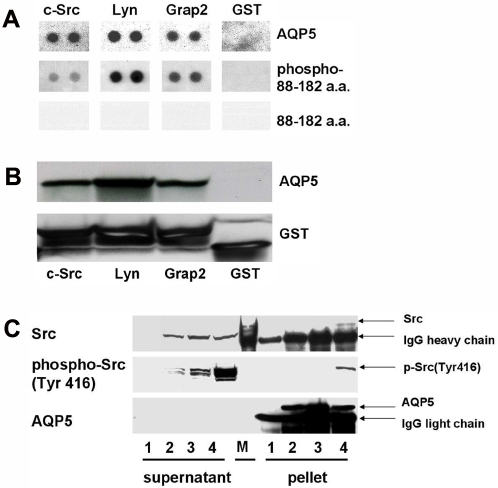
The aquaporins (AQP) are water channel proteins playing a major role in transcellular and transepithelial water movement. Recently, the role of AQPs in human carcinogenesis has become an area of great interest. Here, by immunohistochemistry (IHC), we have found an expression of AQP5 protein in 35.3% (IHC-score: > or = 1, 144/408) of the resected NSCLC tissue samples. Cases with AQP5-positive status (IHC-score: > or = 2) displayed a higher rate of tumor recurrence than negative ones in NSCLC (54.7% vs. 35.1%, p = 0.005) and worse disease-free survival (p = 0.033; OR = 1.52; 95%CI: 1.04-2.23). Further in vitro invasion assay using BEAS-2B and NIH3T3 cells stably transfected with overexpression constructs for full length wild-type AQP5 (AQP5) and its two mutants, N185D which blocks membrane trafficking and S156A which blocks phosphorylation on Ser156, showed that AQP5 induced cell invasions while both mutants did not. In BEAS-2B cells, the expression of AQP5 caused a spindle-like and fibroblastic morphologic change and losses of cell-cell contacts and cell polarity. Only cells with AQP5, not either of two mutants, exhibited a loss of epithelial cell markers and a gain of mesenchymal cell markers. In a human SH3-domains protein array, cellular extracts from BEAS-2B with AQP5 showed a robust binding activity to SH3-domains of the c-Src, Lyn, and Grap2 C-terminal. Furthermore, in immunoprecipitation assay, activated c-Src, phosphorylated on Tyr416, showed a stronger binding activity to cellular extracts from BEAS-2B with AQP5 compared with N185D or S156A mutant. Fluorescence in situ hybridization (FISH) analysis failed to show evidence of genomic amplification, suggesting AQP5 expression as a secondary event. Based on these clinical and molecular observations, we conclude that AQP5, through its phosphorylation on Ser156 and subsequent interaction with c-Src, plays an important role in NSCLC invasion and, therefore, may provide a unique opportunity for developing a novel therapeutic target as well as a prognostic marker in NSCLC.


Conflict of interest statement

Competing Interests: D.S. is a paid consultant to Cangen Biotechnology.

Figures

Figure 1
Figure 1. Clinical importance of AQP5 in non small cell lung cancer. (A) Immunohistochemistry of AQP5.
Photomicrographs of AQP5 immunostaining in arrayed non small cell lung cancer (NSCLC) tissues. Examples of weak (+1), moderate (+2), and strong (+3) immunoreactivity in NSCLCs (a to c, respectively) Original magnification ×200. (B) Kaplan Meier Curve in NSCLC patients. AQP5-positive cases displayed a less favorable disease-free survival rate (log rank p = 0.011) than AQP5-negative cases.
Figure 2
Figure 2. Invasion assay with stable cells expressing AQP5 and mutants.
Matrigel invasion assay was performed with each of BEAS-2B cells (A, C) and NIH3T3 cells (B, D) which express AQP5 or each of two mutants. AQP5 induced cell migration and invasion in BEAS-2B, immortalized human bronchial epithelial cells as well as NIH3T3 cells. In contrast, both of the N185D mutant (impaired membrane trafficking) and the S156A mutant (impaired phosphorylation at Ser 156 by PKA) do not induce cell migration and invasion in either BEAS-2B cells or NIH3T3 cells as compared to Mock.
Figure 3
Figure 3. The Morphologies of stable cells expressing AQP5 and mutants.
The morphologies of the BEAS-2B human bronchial epithelial cells expressing the mock vector FLAG or FLAG/AQP5 were revealed by phase contrast microscopy. BEAS-2B cells maintained highly organized cell-to-cell adhesion and cell polarity as a typical epithelial morphology. The expression of AQP5 caused a spindle-like and fibroblastic morphology and loss of cell-cell contacts and cell polarity. Scale bars, 50 µm.
Figure 4
Figure 4. Immunoblotting analysis of epithelial-mesenchymal transition markers in stable cells expressing AQP5 and mutants.
Expression of epithelial proteins including E-cadherin, α-catenin and γ-catenin, and mesenchymal proteins including fibronectin and vimentin was examined by immunoblotting in the four separate BEAS-2B cells, each of which carries overexpression construct for AQP5 (clone #1 and #2), N185D, S156A and Mock. Only cells with AQP5 exhibited a loss of epithelial markers and a gain of mesenchymal cell markers. 1, AQP5 clone#1; 2, AQP5 clone#2; 3, N185D; 4, S156A; 5, Mock based on BEAS-2B cell lines.
Figure 5
Figure 5. Interaction between AQP5 and the Src family molecules.
(A) A human SH3 domains protein array was used to examine the potential binding activities of several SH3 domain-containing proteins to AQP5. Not only AQP5, but also AQP5 segment containing a phosphorylated Loop D (88 aa through 182 aa) were found to bind to the SH3 domain of the c-Src, the Lyn, and the Grap2 protein. The AQP5 segment containing an unphosphorylated Loop D, however, was unable to bind to the SH3 domain of any of these proteins. (B) GST pull-down assay was performed with three different GST fusion proteins: a c-Src SH3 domain, a Lyn SH3 domain, and a C-terminal Grap2 SH3 domain protein. Cells stably expressing AQP5 in human bronchial epithelial cells, BEAS-2B, revealed a strong interaction with the c-Src SH3 domain protein, the Lyn SH3 domain protein and the C-terminal Grap2 SH3 domain protein. (C) AQP5 is co-immunoprecipitated with c-Src in the BEAS-2B cells stably transfected with AQP5 expression construct. Furthermore, only an activated form of c-Src, phosphorylated on Tyr 416, is co-immunoprecipitated with AQP5. 1, Mock; 2, N185D; 3, S156A; 4, AQP5; M, molecular weight marker.

References

    1. King LS, Agre P. Pathophysiology of the aquaporin water channels. Annu Rev Physiol. 1996;58:619. - PubMed
    1. Verkman AS, van Hoek AN, Ma T, Frigeri A, Skach WR, et al. Water transport across mammalian cell membranes. Am J Physiol. 1996;270:C12–30. - PubMed
    1. Vogelstein B, Kinzler KW. Cancer genes and the pathways they control. Nat Med. 2004;10:789–99. - PubMed
    1. Saadoun S, Papadopoulos MC, Davies DC, Bell BA, Krishna S. Increased aquaporin 1 water channel expression in human brain tumours. Br J Cancer. 2002;87:621–3. - PMC - PubMed
    1. Moon C, Soria JC, Jang SJ, Lee J, Hoque M, et al. Involvement of aquaporins in colorectal carcinogenesis. Oncogene. 2003;22:6699–703. - PubMed

Publication types