

Efficient array-based identification of novel cardiac genes through differentiation of mouse ESCs
- PMID: 18478100
- PMCID: PMC2364653
- DOI: 10.1371/journal.pone.0002176
Efficient array-based identification of novel cardiac genes through differentiation of mouse ESCs
Abstract
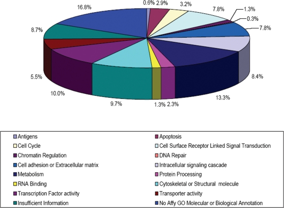
Remarkably, although cardiac disease accounts for the largest proportion of adult mortality and morbidity in the industrialized world, the genetic programs controlling early cardiogenesis are largely incompletely understood. To better understand this process, we set out to identify genes whose expression is enriched within early cardiac fated populations, obtaining the transcriptional signatures of mouse embryonic stem cells (mESCs) at defined intervals during their differentiation along a cardiac path. We compared the RNA profiles of cardiac precursors cells (CPCs) with time-matched non-CPCs and undifferentiated mESCs, using a transgenic mESC line harboring an Nkx2-5 cardiac-specific regulatory sequence driving green fluorescent protein (GFP) to facilitate selection of CPCs. We identify 176 transcripts that are significantly elevated in their abundance within CPCs compared with other assayed populations, predicting that they will likely play a role in cardiogenesis. Of note, approximately 24% (43/176) of the cardiogenic candidate transcripts have known roles in cardiac function or development. Importantly, we evaluated the biological relevance of a significant subset 31/133 (23%) of the remaining candidate genes by in situ hybridization at multiple time points during development (embryonic day, E7.5-9.5) and report that all were expressed in key cardiac structures during cardiogenesis. Furthermore 9/31, of which many were previously uncharacterized, were detected as early as the formation of the cardiac crescent. These data demonstrate the potential power of integrating genomic approaches with mESC differentiation to illuminate developmental processes, and provides a valuable resource that may be mined to further elucidate the genetic programs underlying cardiogenesis.
Conflict of interest statement
Figures


Similar articles
-
Mouse ES cell-derived cardiac precursor cells are multipotent and facilitate identification of novel cardiac genes.J Clin Invest. 2008 Mar;118(3):894-903. doi: 10.1172/JCI33942. J Clin Invest. 2008. PMID: 18246200 Free PMC article.
-
Mouse embryonic stem cells irradiated with γ-rays differentiate into cardiomyocytes but with altered contractile properties.Mutat Res. 2013 Aug 30;756(1-2):37-45. doi: 10.1016/j.mrgentox.2013.06.007. Epub 2013 Jun 20. Mutat Res. 2013. PMID: 23792212
-
Identification of cardiovascular lineage descendants at single-cell resolution.Development. 2015 Mar 1;142(5):846-57. doi: 10.1242/dev.116897. Epub 2015 Jan 29. Development. 2015. PMID: 25633351 Free PMC article.
-
Gene expression profiling reveals the heterogeneous transcriptional activity of Oct3/4 and its possible interaction with Gli2 in mouse embryonic stem cells.Genomics. 2013 Nov-Dec;102(5-6):456-67. doi: 10.1016/j.ygeno.2013.09.004. Epub 2013 Oct 8. Genomics. 2013. PMID: 24121003
-
Efficient capture of cardiogenesis-associated genes expressed in ES cells.Biochem Biophys Res Commun. 2007 Mar 30;355(1):47-53. doi: 10.1016/j.bbrc.2007.01.109. Epub 2007 Jan 30. Biochem Biophys Res Commun. 2007. PMID: 17286962
Cited by
-
A review of human pluripotent stem cell-derived cardiomyocytes for high-throughput drug discovery, cardiotoxicity screening, and publication standards.J Cardiovasc Transl Res. 2013 Feb;6(1):22-30. doi: 10.1007/s12265-012-9423-2. Epub 2012 Nov 15. J Cardiovasc Transl Res. 2013. PMID: 23229562 Free PMC article. Review.
-
The Involvement of Long Non-coding RNA and Messenger RNA Based Molecular Networks and Pathways in the Subacute Phase of Traumatic Brain Injury in Adult Mice.Front Neuroinform. 2022 Mar 4;16:794342. doi: 10.3389/fninf.2022.794342. eCollection 2022. Front Neuroinform. 2022. PMID: 35311004 Free PMC article.
-
IGF-2/IGF-1R signaling has distinct effects on Sox1, Irx3, and Six3 expressions during ES cell derived-neuroectoderm development in vitro.In Vitro Cell Dev Biol Anim. 2016 May;52(5):607-15. doi: 10.1007/s11626-016-0012-6. Epub 2016 Mar 8. In Vitro Cell Dev Biol Anim. 2016. PMID: 26956358
-
Expression and mutation analyses implicate ARHGAP29 as the etiologic gene for the cleft lip with or without cleft palate locus identified by genome-wide association on chromosome 1p22.Birth Defects Res A Clin Mol Teratol. 2012 Nov;94(11):934-42. doi: 10.1002/bdra.23076. Epub 2012 Sep 24. Birth Defects Res A Clin Mol Teratol. 2012. PMID: 23008150 Free PMC article.
-
Transmembrane protein 108 involves in adult neurogenesis in the hippocampal dentate gyrus.Cell Biosci. 2019 Jan 11;9:9. doi: 10.1186/s13578-019-0272-4. eCollection 2019. Cell Biosci. 2019. PMID: 30651970 Free PMC article.
References
-
- Harvey RP. Patterning the vertebrate heart. Nat Rev Genet. 2002;3:544–556. - PubMed
-
- Tam PP, Parameswaran M, Kinder SJ, Weinberger RP. The allocation of epiblast cells to the embryonic heart and other mesodermal lineages: the role of ingression and tissue movement during gastrulation. Development. 1997;124:1631–1642. - PubMed
-
- Kelly RG, Brown NA, Buckingham ME. The arterial pole of the mouse heart forms from Fgf10-expressing cells in pharyngeal mesoderm. Dev Cell. 2001;1:435–440. - PubMed
-
- Abu-Issa R, Waldo K, Kirby ML. Heart fields: one, two or more? Dev Biol. 2004;272:281–285. - PubMed
Publication types
MeSH terms
Grants and funding
LinkOut - more resources
Full Text Sources
Molecular Biology Databases
