. 2008 Jul;10(4):535-41.
doi: 10.1111/j.1745-7262.2008.00410.x.

Characterization of nucleohistone and nucleoprotamine components in the mature human sperm nucleus

Affiliations

Characterization of nucleohistone and nucleoprotamine components in the mature human sperm nucleus

Yan Li et al. Asian J Androl. 2008 Jul.

Abstract

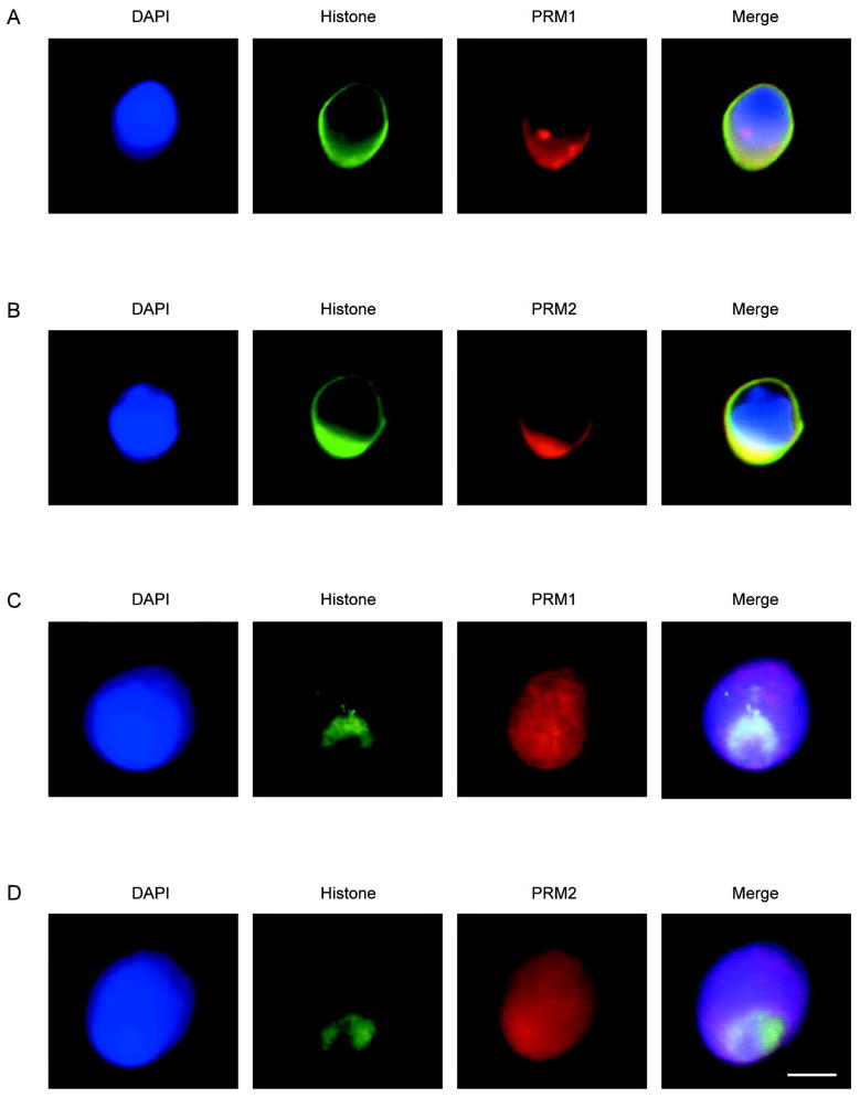
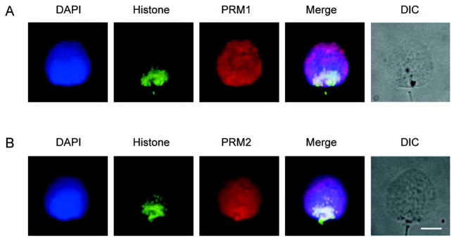
Aim: To simultaneously determine the localization of histones and protamines within human sperm nuclei.

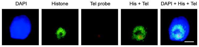
Methods: Immunofluorescence of the core histones and protamines and fluorescence in situ hybridization of the telomere region of chromosome 16 was assessed in decondensed human sperm nuclei.

Results: Immunofluorescent localization of histones, protamine 1 (PRM1) and protamine 2 (PRM2) along with fluorescence in situ hybridization localization of chromosome 16 telomeric sequences revealed a discrete distribution in sperm nuclei. Histones localized to the posterior ring region (i.e. the sperm nuclear annulus), whereas PRM1 and PRM2 appeared to be dispersed throughout the entire nucleus.

Conclusion: The co-localization of the human core sperm histones with the telomeric regions of chromosome 16 is consistent with the reorganization of specific non-protamine regions into a less compacted state.


Figures

Figure 1
Figure 1
Immunofluorescent localization of the core histones, protamine 1 (PRM1) and protamine 2 (PRM2) in human sperm nuclei. (A): Dual histone and PRM1 immunofluorescence in decondensed nuclei. (B): Dual histone and PRM2 immunofluorescence in decondensed nuclei. (C): Immunofluorescent colocalization of core histones with PRM1 in decondensed and permeablized nuclei. (D) Immunofluorescent colocalization of histones with PRM2 in decondensed and permeablized nuclei. DNA stained with blue 4′-6-diamidino-2-phenylindole (DAPI), histones green, with Alexa Fluor 488-conjugated streptavidin and protamines red with CY5-conjugated anti-mouse antibody. The merged pseudo-color image is shown in the right most panel. Scale bar=5 μm. of a histone-enriched territory well apart from the PRM1 and PRM2 regions distributed throughout the nucleus. The question arises, what does this unique packaging reflect?
Figure 2
Figure 2
Immunofluorescent colocalization of the core histones with protamine 1 (PRM1) and protamine 2 (PRM2) in permeablized and decondensed human sperm cells. (A): Immunofluorescent colocalization of histones with anti-core histones antibody plus PRM1 with anti-protamine 1. (B): Immunofluorescent colocalization of histones with anti-core histones antibody plus PRM2 with anti-protamine 2. The corresponding grey-scale DIC image of the intact permeablized cells is shown. DNA stained with blue 4′-6-diamidino-2-phenylindole (DAPI), histones green, with Alexa Fluor 488-conjugated streptavidin and protamines red with CY5-conjugated goat anti-mouse antibody. The merged pseudo-color image is shown in the right most panel. Scale bar=5 μm. Note that core the histones localize towards the post acrosomal posterior ring region end while PRM1 and PRM2 occupy the entire nucleus.
Figure 3
Figure 3
Distribution of the core histones and the telomeric region of human chromosome 16 within the permeablized and decondensed sperm nucleus. The fluorescence in situ hybridization signal (FISH) shows that the human chromosome 16 telomeric region is located within an immunofluorescent core histone rich region. The relative location of the biotin-labeled human chromosome 16 telomeric region is indicated in red with streptavidin Alexa Fluor 594 whereas the core histone region marked by green with Alexa Fluor 488. The merged pseudo-color image is shown in the right most panel. Scale bar=5 μm. DAPI, 4′-6-diamidino-2-phenylindole; Tel, telomere; His, histones.

References

    1. Holstein AF, Schulze W, Davidoff M. Understanding spermatogenesis is a prerequisite for treatment. Reprod Biol Endocrinol. 2003;1:107. - PMC - PubMed
    1. Krawetz SA. Paternal contribution: new insights and future challenges. Nat Rev Genet. 2005;6:633–42. - PubMed
    1. Rousseaux S, Caron C, Govin J, Lestrat C, Faure AK, Khochbin S. Establishment of male-specific epigenetic information. Gene. 2005;345:139–53. - PubMed
    1. Berger SL. Histone modifications in transcriptional regulation. Curr Opin Genet Dev. 2002;12:142–8. - PubMed
    1. Govin J, Caron C, Lestrat C, Rousseaux S, Khochbin S. The role of histones in chromatin remodelling during mammalian spermiogenesis. Eur J Biochem. 2004;271:3459–69. - PubMed

Publication types