Review
. 2008 Jun;30(6):530-41.
doi: 10.1002/bies.20767.

The origin and evolution of the neural crest

Affiliations
Review

The origin and evolution of the neural crest

Philip C J Donoghue et al. Bioessays. 2008 Jun.

Abstract

Many of the features that distinguish the vertebrates from other chordates are derived from the neural crest, and it has long been argued that the emergence of this multipotent embryonic population was a key innovation underpinning vertebrate evolution. More recently, however, a number of studies have suggested that the evolution of the neural crest was less sudden than previously believed. This has exposed the fact that neural crest, as evidenced by its repertoire of derivative cell types, has evolved through vertebrate evolution. In this light, attempts to derive a typological definition of neural crest, in terms of molecular signatures or networks, are unfounded. We propose a less restrictive, embryological definition of this cell type that facilitates, rather than precludes, investigating the evolution of neural crest. While the evolutionary origin of neural crest has attracted much attention, its subsequent evolution has received almost no attention and yet it is more readily open to experimental investigation and has greater relevance to understanding vertebrate evolution. Finally, we provide a brief outline of how the evolutionary emergence of neural crest potentiality may have proceeded, and how it may be investigated.


Figures
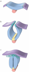
Figure 1
Figure 1
The principal derivative cell types of neural crest. The major derivatives of neural crest cells are schematically illustrated here, grouped according to whether they are classified as ectomesenchymal or non-ectomesenchymal.
Figure 2
Figure 2
Stages in the formation of the neural crest. A) At the neural plate stage the non-neural ectoderm signals to the neural plate, primarily through the actions of Bmp’s and wnt’s, to specify the neural plate border region. The notochord underlies the midline of the neural plate. B) As neurulation progresses, the neural plate infolds and the neural plate border rises up and forms the neural folds. At the ventral midline of the neural plate, the floor plate is induced to form by the notochord. The floor plate and notochord will act to pattern the ventral neural tube. C) Following apposition of the neural folds and their fusion across the midline, the neural tube is forms. The neural crest cells emigrate from the dorsal most region of the neural tube, which is derived from the neural folds. This dorsal territory will also generate the roof plate, and this structure will act to pattern the dorsal neural tube.
Figure 3
Figure 3
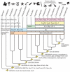
The evolutionary assembly of the neural crest gene regulatory network (GRN). Across the tips of the tree we have indicated by the extent of the box, enveloping more or less branches, the evolutionary addition of the various elements of the neural crest GRN as identified by Sauka-Spengler and colleagues(16). This clearly shows that although functional integration of neural crest specifiers and effectors into the GRN occurs only in vertebrates, the early patterning and neural plate border specification network components have a much more protracted evolutionary history, extending at least to the last common ancestor of triploblastic bilaterian eumetazoans. The integration of the key signalling molecules and transcription factors into the neural crest GRN should not, however, be confused with the evolutionary origin of these factors, the majority of which were present in the last common ancestor of metazoans, as indicated by their point of origin in the internal branches of the tree. Finally, the apparent point of evolutionary origin of major neural crest cell phenotypes is also indicated on the internal branches of the tree.
Figure 4
Figure 4
Hierarchical relationships between principal categories of neural crest cell-type derivatives based on a cladistic analysis of their characteristics, undertaken by Vickaryous and Hall(52). The analysis is based on a character matrix that encompasses aspects of cell phenotype, behaviour and labelling, and was subjected to both phenetic cluster analysis and cladistic parsimony analysis; only the results of the parsimony analysis are presented here. The hierarchy bears out the dichotomy between ectomesenchymal (e) and non-ectomesenchymal (n) fates.

References

    1. Gans C, Northcutt RG. Neural crest and the origin of the vertebrates: a new head. Science. 1983;220:268–274. - PubMed
    1. Northcutt RG, Gans C. The genesis of neural crest and epidermal placodes: a reinterpretation of vertebrate origins. Quarterly Review of Biology. 1983;58:1–28. - PubMed
    1. Bernhardt RR, Chitnis AB, Lindamer L, Kuwada JY. Identification of spinal neurons in the embryonic and larval zebrafish. Journal of Comparative Neurology. 1990;302:603–616. - PubMed
    1. Fritzsch B, Northcutt RG. Cranial and spinal nerve organization in Amphioxus and lampreys: evidence for an ancestral craniate pattern. Acta Anatomica. 1993;148:96–109. - PubMed
    1. Cornell RA, Eisen JS. Delta signaling mediates segregation of neural crest and spinal sensory neurons from zebrafish lateral neural plate. Development. 2000;127:2873–2882. - PubMed

Publication types