. 2008 Nov;49(11):1893-907.
doi: 10.1111/j.1528-1167.2008.01656.x. Epub 2008 May 9.

Interictal high-frequency oscillations (80-500 Hz) are an indicator of seizure onset areas independent of spikes in the human epileptic brain

Affiliations

Interictal high-frequency oscillations (80-500 Hz) are an indicator of seizure onset areas independent of spikes in the human epileptic brain

Julia Jacobs et al. Epilepsia. 2008 Nov.

Abstract

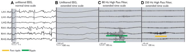
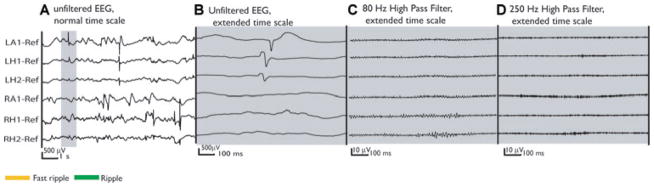
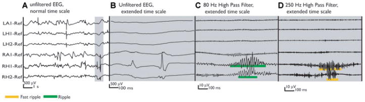
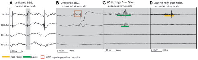
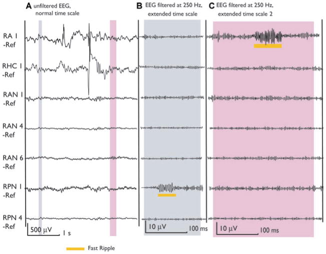
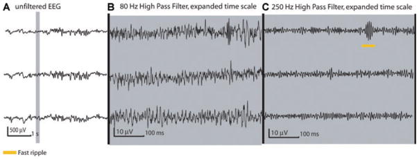
Purpose: High-frequency oscillations (HFOs) known as ripples (80-250 Hz) and fast ripples (250-500 Hz) can be recorded from macroelectrodes inserted in patients with intractable focal epilepsy. They are most likely linked to epileptogenesis and have been found in the seizure onset zone (SOZ) of human ictal and interictal recordings. HFOs occur frequently at the time of interictal spikes, but were also found independently. This study analyses the relationship between spikes and HFOs and the occurrence of HFOs in nonspiking channels.

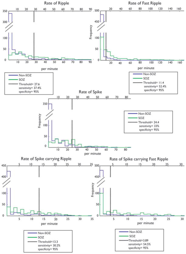
Methods: Intracerebral EEGs of 10 patients with intractable focal epilepsy were studied using macroelectrodes. Rates of HFOs within and outside spikes, the overlap between events, event durations, and the percentage of spikes carrying HFOs were calculated and compared according to anatomical localization, spiking activity, and relationship to the SOZ.

Results: HFOs were found in all patients, significantly more within mesial temporal lobe structures than in neocortex. HFOs could be seen in spiking as well as nonspiking channels in all structures. Rates and durations of HFOs were significantly higher in the SOZ than outside. It was possible to establish a rate of HFOs to identify the SOZ with better sensitivity and specificity than with the rate of spikes.

Discussion: HFOs occurred to a large extent independently of spikes. They are most frequent in mesial temporal structures. They are prominent in the SOZ and provide additional information on epileptogenicity independently of spikes. It was possible to identify the SOZ with a high specificity by looking at only 10 min of HFO activity.


Conflict of interest statement

Conflict of interest: We confirm that we have read the Journal’s position on issues involved in ethical publication and affirm that this report is consistent with those guidelines. The authors have no professional or financial affiliations that might be perceived as having biased the presentation.

Figures

Figure 1
Figure 1
Patient 5 presented with interictal and ictal signs of bitemporal epilepsy on scalp EEG. He was implanted bitemporally aiming at the amygdala (LA, RA) and hippocampus (LH, RH). The MRI revealed a malrotation of the right hippocampus. All seizures originated from the right mesial temporal structures, where the highest rates of HFOs were found. (A) The unfiltered EEG shows frequent spikes in both mesial TL structures. The thin gray section in A is expanded in time and amplitude in B, C, and D demonstrating the cooccurrence of ripples and fast ripples outside spikes in channel RH1. Additionally, a ripple is seen in channel RH2. Note that C and D have a much higher gain than A and B. Epilepsia © ILAE
Figure 2
Figure 2
Same unfiltered EEG as in Fig. 1 (A). This time the selection includes two spikes over LA1 and LH1. Both spikes lied outside of the seizure onset zone and in the healthier mesial temporal lobe. The expanded sections of the unfiltered EEG (B) and filtered EEG (C, D) show no high-frequency oscillation during this time period. Epilepsia © ILAE
Figure 3
Figure 3
Same unfiltered EEG segment as in Figs. 1(A) and 2 (A). The gray selection shows two spikes recorded simultaneously at RH1 and RH2 in the SOZ. No oscillation is visible in the spike at RH1 in the extended unfiltered EEG (B), while the spike in RH2 shows a very short and small oscillation. The filtered EEG segments clearly reveal a ripple and fast ripple oscillation during these spikes (C, D). Compared with Fig. 2, this demonstrates that spikes which look similar in the unfiltered EEG might differ in whether they carry high-frequency oscillations or not. This difference is only visible after filtering. Epilepsia © ILAE
Figure 4
Figure 4
Patient 1 was implanted bilaterally in the mesial temporal structures. Bilateral interictal spikes were observed, but all recorded seizures started in the left mesial temporal structures. The unfiltered EEG shows three spikes over LH1 and LH2 (A). The selected gray segment shows the extended unfiltered EEG of one spike, which in LH1 showed a clear oscillation riding on the spike (B). When the EEG is filtered, ripples and fast ripples are seen at the time of the spike in LH1 and LH2 channels, but much more prominently at LH1 (C, D). Epilepsia © ILAE
Figure 5
Figure 5
The MRI of patient 6 showed two heterotopic nodules located in the right trigonal area. Two contacts are inside these nodules (RAN1, RPN1). Ictal and interictal discharges were seen on the scalp over the right temporal lobe and the patient was implanted in the right hippocampus (RHC) and Amygdala (RA). Contacts RA1 and RHC1 were in the SOZ. This figure shows two selections (gray) of the unfiltered EEG. The first demonstrates a fast ripple outside spikes in a nonspiking channel (RPN1). This channel showed only fast ripples and no other epileptiform activity and none of the patient’s seizure was generated in this lesion. The second selection shows a fast ripple inside SOZ but outside a spike. It is important to notice that most of the HFOs in neocortical areas and outside spikes were shorter and of lower amplitude than those within spikes. This is visible when comparing with the HFOs in Fig. 1 inside the mesial temporal lobe or with the ones in Figs. 3 and 4 cooccurring with spikes. Epilepsia © ILAE
Figure 6
Figure 6
[Correction added after online publication July 11, 2008: The gain for the EEG traces in part B of Figure 6 was incorrect in the original publication. A corrected figure is provided above.] Patient 10 presented with frontal lobe seizures and a focal cortical dysplasia in the left second frontal gyrus. Electrode LS was implanted inside this area with contacts 1–5 in the lesion. All of these channels were spiking and we selected the three displayed in this figure for analysis. The selection shows a fast ripple (at LS1) occurring independently of spiking or ripple activity. Even though all of the patient’s seizures were generated within these channels, the rate of HFOs was generally low compared to that observed in the mesial temporal structures in other patients. As can be seen here the fast ripples were shorter and more discreet (compare with Fig. 1). Epilepsia © ILAE
Figure 7
Figure 7
Histograms comparing channels inside and outside the SOZ. The five histograms show the rates of spikes, ripples, fast ripples, spikes with ripples and spikes with fast ripples. Green indicates rates in SOZ and blue outside SOZ. The thresholds for 95% specificity are shown as black vertical lines. If the SOZ was not known and if a rate above this threshold was selected to separate SOZ from non-SOZ channels, the indicated sensitivity would result. The rate of fast ripples and of spikes with fast ripples showed the highest sensitivity in indicating the SOZ. Epilepsia © ILAE

References

    1. Bautista RE, Cobbs MA, Spencer DD, Spencer SS. Prediction of surgical outcome by interictal epileptiform abnormalities during intracranial EEG monitoring in patients with extrahippocampal seizures. Epilepsia. 1999;40(7):880–890. - PubMed
    1. Benini R, D’Antuono M, Pralong E, Avoli M. Involvement of amygdala networks in epileptiform synchronization in vitro. Neuroscience. 2003;120:75–84. - PubMed
    1. Bragin A, Engel J, Jr, Wilson CL, Fried I, Mathern GW. Hippocampal and entorhinal cortex high-frequency oscillations (100–500 Hz) in human epileptic brain and in kainic acid-treated rats with chronic seizures. Epilepsia. 1999;40:127–137. - PubMed
    1. Bragin A, Wilson CL, Staba RJ, Reddick M, Fried I, Engel J., Jr Interictal high-frequency oscillations (80–500 Hz) in the human epileptic brain: entorhinal cortex. Ann Neurol. 2002;52:407–415. - PubMed
    1. Bragin A, Wilson CL, Engel J. Spatial stability over time of brain areas generating fast ripples in the epileptic rat. Epilepsia. 2003;44:1233–1237. - PubMed

Publication types