

Directed evolution of a biterminal bacterial display scaffold enhances the display of diverse peptides
- PMID: 18480093
- PMCID: PMC2427320
- DOI: 10.1093/protein/gzn020
Directed evolution of a biterminal bacterial display scaffold enhances the display of diverse peptides
Abstract
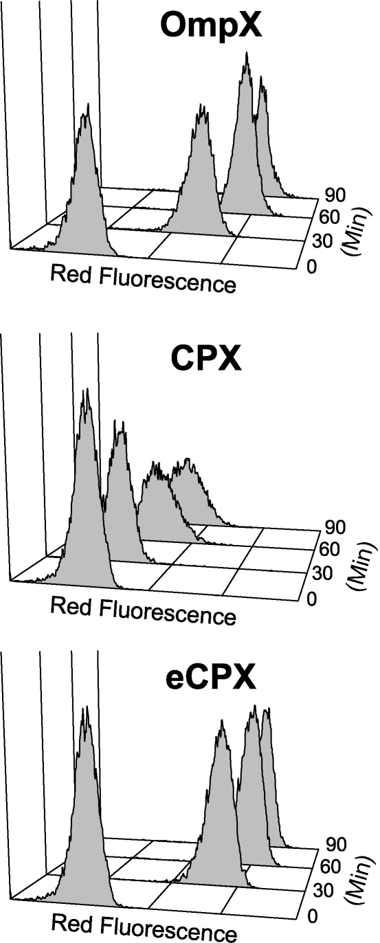
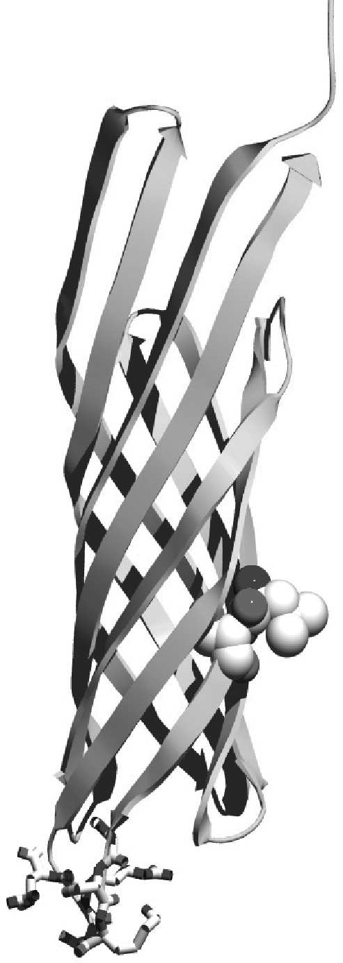
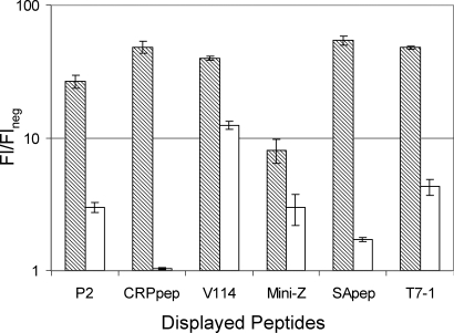
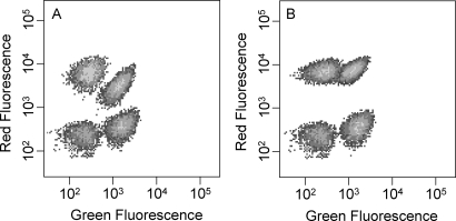
Bacterial cell-surface display systems coupled with quantitative screening methods offer the potential to expand protein engineering capabilities. To more fully exploit this potential, a unique bacterial surface display scaffold was engineered to display peptides more efficiently from the surface exposed C- and N-termini of a circularly permuted outer membrane protein. Using directed evolution, efficient membrane localization of a circularly permuted OmpX (CPX) display scaffold was rescued, thereby improving the presentation of diverse passenger peptides on the cell surface. Random and targeted mutagenesis directed towards linkers joining the native N- and C-termini of OmpX coupled with screening by FACS yielded an enhanced CPX (eCPX) variant which localized to the outer membrane as efficiently as the non-permuted parent. Interestingly, enhancing substitutions coincided with a C-terminal motif conserved in outer membrane proteins. Surface localization of various passenger peptides and mini-proteins was expedited using eCPX relative to that achieved with the parent scaffold. The new variant also permitted simultaneous display and labeling of distinct peptides on structurally adjacent C- and N-termini, thus enabling display level normalization during library screening and the display of bidentate or dimeric peptides. Consequently, the evolved scaffold, eCPX, expands the range of applications for bacterial display. Finally, this approach provides a route to improve the performance of cell-surface display vectors for protein engineering and design.
Figures
References
-
- Becker S., Theile S., Heppeler N., Michalczyk A., Wentzel A., Wilhelm S., Jaeger K.E., Kolmar H. FEBS Lett. 2005;579:1177–1182. - PubMed
-
- Bessette P.H., Rice J.J., Daugherty P.S. Protein Eng. Des. Sel. 2004;17:731–739. - PubMed
-
- Boder E.T., Wittrup K.D. Nat. Biotechnol. 1997;15:553–557. - PubMed
-
- Bos M.P., Tommassen J. Curr. Opin. Microbiol. 2004;7:610–616. - PubMed
Publication types
MeSH terms
Substances
LinkOut - more resources
Full Text Sources
Other Literature Sources
Research Materials
